诊断学理论与实践 ›› 2021, Vol. 20 ›› Issue (02): 161-167.doi: 10.16150/j.1671-2870.2021.02.008
钱乐, 姜美娇, 杨伯文, 陈慧( )
)
收稿日期:2021-01-29
出版日期:2021-04-25
发布日期:2022-06-28
通讯作者:
陈慧
E-mail:ch11516@rjh.com.cn
基金资助:
QIAN Le, JIANG Meijiao, YANG Bowen, CHEN Hui()
Received:2021-01-29
Online:2021-04-25
Published:2022-06-28
Contact:
CHEN Hui
E-mail:ch11516@rjh.com.cn
摘要:
目的:探讨卵巢囊性腺纤维瘤(cystadenofibroma,CAF)和卵巢腺纤维瘤(adenofibroma,AF)的超声图像特征。方法:回顾性分析经术后病理检查证实的50例卵巢CAF和7例卵巢AF患者的超声声像图特征,并评估超声的诊断价值。结果:根据病理结果,50例CAF中,良性为46例,共47个肿块;交界性为4例,均为单发,7例AF均为良性单发病灶。卵巢CAF和AF的病理分型以浆液性为主(53/58,91.4%)。将交界性肿瘤归入恶性肿瘤,术前超声检查鉴别CAF及AF肿块良恶性的灵敏度为100.0%[95%置信区间(confidential interval,95%CI)为39.8%~100.0%],特异度为72.2%(95%CI为58.4%~83.5%),准确率为74.1%。超声图像上,良性CAF多表现为形态规则(43/47,91.5%)、边界清晰(46/47,97.9%)的单房囊实性肿块(25/47,53.2%),且多数肿块附壁有规则的乳头状突起(32/47,68.1%),突起内无或少量血流信号(31/32,96.9%)。交界性CAF的超声表现均有实性部分或乳头状突起,内部血供程度为少量至丰富不等。与良性CAF相比,交界性CAF实性部分或乳头状突起的最大径更大[最大径,19.5(18.8~26.3) mm比8.0(7.0~13.0) mm,P=0.002],实性部分或乳头状突起与肿块最大径比值更大(0.35比0.15,P=0.033),内有血供者更多(4/4比10/35,P=0.012)。AF超声图像多表现为形态规则(6/7)、边界清晰(5/7)且后方伴有回声衰减的实性肿块(4/7),其实性部分内呈乏血供表现(6/6)。结论:超声诊断CAF及AF的效能一般。良性CAF多表现为附壁有乏血供乳头状突起的囊实性肿块;交界性CAF多表现为有不同程度血供的实性或乳头状突起的肿块;而AF多表现为后方伴回声衰减、无血供的实性肿块。肿块在超声图像上表现为实性成分多和血供程度丰富,多提示交界性CAF。
中图分类号:
钱乐, 姜美娇, 杨伯文, 陈慧. 卵巢囊性腺纤维瘤和卵巢腺纤维瘤的超声诊断效能及图像特征分析[J]. 诊断学理论与实践, 2021, 20(02): 161-167.
QIAN Le, JIANG Meijiao, YANG Bowen, CHEN Hui. The ultrasonic features and diagnostic performance of ultrasound for ovarian cystadenofibroma and adenofibroma[J]. Journal of Diagnostics Concepts & Practice, 2021, 20(02): 161-167.
表1
卵巢CAF和AF的临床特征[n(%)]
| 特征 | 良性CAF (46例) | AF (7例) | 交界性CAF (4例) |
|---|---|---|---|
| 年龄(岁) | 48(33.3~58.0) | 48(45.0~56.0) | 38.5(29.0~56.0) |
| 既往卵巢手术史 | 3(6.5%) | 1 | 0 |
| 卵巢癌家族史或个人史 | 0/0 | 0/0 | 0/0 |
| 已绝经/未绝经 | 19/27 | 3/4 | 1/3 |
| 临床症状 | |||
| 腹痛、腹胀、腹围增大 | 10(21.7%) | 0 | 0 |
| 腹部触及包块 | 10(21.7%) | 2 | 0 |
| 阴道异常出血 | 2(4.3%) | 1 | 0 |
| 体重明显改变 | 2(4.3%) | 1 | 0 |
| 无明显症状 | 22(47.8%) | 3 | 4 |
| 肿瘤指标 CA12-5>35 U/mL [n(%)] | 5(10.9%) | 0 | 1 |
| CA12-5(U/mL) | 12.3(10.1~18.7) | 9.7(9.1~10.8) | 21.2(15.7~205.1) |
| HE4>60.5 U/mL [n(%)] | 7(15.2%) | 0 | 0 |
| HE4(U/mL) | 38.0(32.9~47.1) | 44.0(40.1~46.7) | 41.2(36.0~43.1) |
表2
卵巢CAF和AF的术前评估及手术病理分型[n(%)]
| 方法 | 良性CAF (n=47) | AF (n=7) | 交界性CAF(n=4) |
|---|---|---|---|
| 超声术前评估 | |||
| 良性 | 32(68.1%) | 7 | 0 |
| 囊腺瘤或单纯性囊肿 | 20(42.6%) | 3 | / |
| 系膜囊肿 | 6(12.8%) | 0 | / |
| 内膜囊肿 | 3(6.4%) | 0 | / |
| AF | 1(2.1%) | 0 | / |
| 卵泡膜-纤维瘤类肿瘤 | 1(2.1%) | 4 | / |
| 畸胎瘤 | 1(2.1%) | 0 | / |
| 恶性或交界性 | 15(31.9%) | 0 | 4 |
| 病理结果 | |||
| 浆液性 | 44(93.6%) | 7 | 2 |
| 黏液性 | 2(4.3%) | 0 | 0 |
| 内膜样 | 1(2.1%) | 0 | 1 |
| 浆-黏液性 | 0(0.0%) | 0 | 1 |
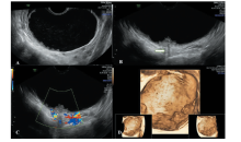
图1
良性浆液性卵巢CAF的超声图像 A:二维灰阶超声显示为一个形态规则、边界清晰的单房囊实性肿块,附壁可见多枚乳头状突起;B:乳头状突起后方可见回声衰减(白色箭头);C:彩色多普勒血流提示乳头内少许点状血流信号;D:三维超声可见附壁多枚乳头状突起。
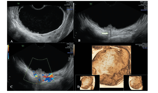
表3
卵巢CAF和AF的超声图像特征及表现[n(%)]
| 特征 | 良性CAF(n=47) | AF(n=7) | 交界性CAF(n=4) |
|---|---|---|---|
| 肿块位置 | |||
| 卵巢内 | 9(19.1%) | 2 | 1 |
| 卵巢旁 | 16(34.0%) | 2 | 1 |
| 无法辨别 | 22(46.8%) | 3 | 2 |
| 肿块最大径(mm) | 47(31~85) | 33(24~70) | 54.5(45~96) |
| 肿块形态规则 | 43(91.5%) | 6 | 2 |
| 肿块边界清晰 | 46(97.9) | 5 | 2 |
| 肿块分类 | |||
| 单房囊性 | 4(8.5%) | 0 | 0 |
| 单房囊实性 | 25(53.2%) | 2 | 1 |
| 实性 | 2(4.3%) | 4 | 1 |
| 多房囊性 | 8(17.0%) | 1 | 0 |
| 多房囊实性 | 8(17.0%) | 0 | 2 |
| 分隔厚度(mm) | 2.3(1.9~3.3) | 1.3 | 1.3/3.7 |
| 实性部分或乳头状突起 | 35(74.5%) | 6 | 4 |
| 实性部分或乳头状突起最大径(mm) | 8.0(7.0~13.0) | 18.5(9.3~30.8) | 19.5(18.8~26.3)a) |
| 实性部分或乳头状突起/肿块最大径比值 | 0.15(0.12~0.25) | 1.00(0.46~1.00) | 0.35(0.30~0.52)b) |
| 乳头状突起 | 32(68.1%) | 2 | 3 |
| 1枚 | 13/32(40.6%) | 0/2 | 0/3 |
| 2枚 | 3/32(9.4%) | 0/2 | 0/3 |
| 3枚 | 5/32(15.6%) | 1/2 | 0/3 |
| ≥4枚 | 11/32(34.4%) | 1/2 | 3/3 |
| 乳头状突起最大径(mm) | 7.5(7.0~11.3) | 8/8 | 6/19/20 |
| 乳头状突起最大径/肿块最大径比值 | 0.15(0.12~0.22) | 0.11,0.29 | 0.11,0.20,0.36 |
| 实性部分或乳头状突起血流评分 | |||
| 1分(无) | 25/35(71.4%) | 6/6 | 0/4c) |
| 2分(少量) | 8/35(22.9%) | 0/6 | 1/4 |
| 3分(中等) | 2/35(5.7%) | 0/6 | 2/4 |
| 4分(丰富) | 0/35(0) | 0/6 | 1/4 |
| 实性部分或乳头状突起后方回声衰减 | 15(31.9%) | 4 | 1 |
| 肿块后方回声增强 | 27(57.4%) | 3 | 2 |

图2
交界性内膜样卵巢CAF的超声图像 A:二维灰阶超声显示形态不规则、边界不清的多房囊实性肿块,分隔上有不规则实性部分,附壁有4枚以上乳头状突起;B:1枚附壁乳头的测量;C:彩色多普勒血流提示,突起及实性部分内见中等血流信号;D:三维超声可见附壁多枚乳头状突起及实性部分。
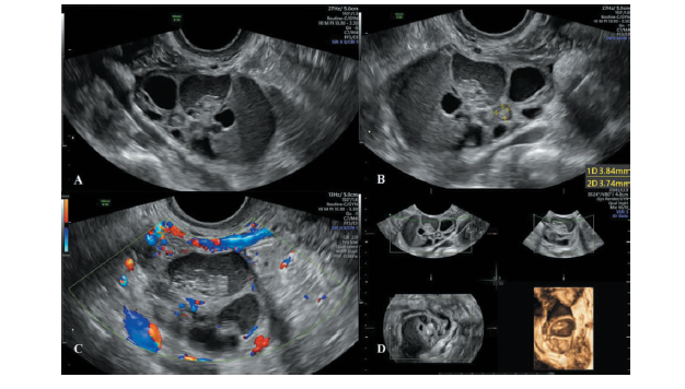

图3
浆液性卵巢AF的超声图像 A:二维灰阶超声提示形态不规则、边界不清晰的实性肿块,肿块后方可见回声衰减;B:彩色多普勒血流提示,肿块内部无明显血流信号。
| [1] |
Taylor EC, Irshaid L, Mathur M. Multimodality imaging approach to ovarian neoplasms with pathologic correlation[J]. Radiographics, 2021, 41(1):289-315.
doi: 10.1148/rg.2021200086 pmid: 33186060 |
| [2] |
Cho SM, Byun JY, Rha SE, et al. CT and MRI findings of cystadenofibromas of the ovary[J]. Eur Radiol, 2004, 14(5):798-804.
doi: 10.1007/s00330-003-2060-z URL |
| [3] |
Czernobilsky B, Borenstein R, Lancet M. Cystadenofibroma of the ovary. A clinicopathologic study of 34 cases and comparison with serous cystadenoma[J]. Cancer, 1974, 34(6):1971-1981.
pmid: 4434327 |
| [4] |
Kozawa E, Inoue K, Takahashi M, et al. Diffusion-weighted MR imaging findings of ovarian adenocarcinofibromas and adenofibromas[J]. Clin Imaging, 2014, 38(4):483-489.
doi: 10.1016/j.clinimag.2014.01.014 URL |
| [5] |
Timmerman D, Valentin L, Bourne TH, et al. Terms, de-finitions and measurements to describe the sonographic features of adnexal tumors: a consensus opinion from the International Ovarian Tumor Analysis(IOTA) Group[J]. Ultrasound Obstet Gynecol, 2000, 16(5):500-505.
doi: 10.1046/j.1469-0705.2000.00287.x URL |
| [6] |
Andreotti RF, Timmerman D, Strachowski LM, et al. ORADS US risk stratification and management system: a consensus guideline from the ACR Ovarian-Adnexal Reporting and Data System Committee[J]. Radiology, 2020, 294(1):168-185.
doi: 10.1148/radiol.2019191150 pmid: 31687921 |
| [7] |
Tang YZ, Liyanage S, Narayanan P, et al. The MRI features of histologically proven ovarian cystadenofibromas-an assessment of the morphological and enhancement patterns[J]. Eur Radiol, 2013, 23(1):48-56.
doi: 10.1007/s00330-012-2568-1 pmid: 22814827 |
| [8] | 潘玉萍, 蔡爱露, 赵丹, 等. 卵巢腺纤维瘤及囊性腺纤维瘤的超声特征[J]. 中国医学影像技术, 2012, 28(9):1699-1701. |
| [9] |
Shimizu S, Okano H, Ishitani K, et al. Ovarian cystadenofibroma with solid nodular components masqueraded as ovarian cancer[J]. Arch Gynecol Obstet, 2009, 279(5):709-711.
doi: 10.1007/s00404-008-0785-2 pmid: 18779972 |
| [10] | 徐健, 梅海炳, 高军, 等. 卵巢浆液性囊性腺纤维瘤MR诊断及病理分析[J]. 医学影像学杂志, 2016, 26(8):1494-1497. |
| [11] |
Virgilio BA, de Blasis I, Sladkevicius P, et al. Imaging in gynecological disease(16): clinical and ultrasound cha-racteristics of serous cystadenofibromas in adnexa[J]. Ultrasound Obstet Gynecol, 2019, 54(6):823-830.
doi: 10.1002/uog.20277 pmid: 30937992 |
| [12] |
Goldstein SR, Timor-Tritsch IE, Monteagudo A, et al. Cystadenofibromas: can transvaginal ultrasound appea-rance reduce some surgical interventions?[J]. J Clin Ultrasound, 2015, 43(6):393-396.
doi: 10.1002/jcu.22241 pmid: 25271400 |
| [13] |
Landolfo C, Valentin L, Franchi D, et al. Differences in ultrasound features of papillations in unilocular-solid adnexal cysts: a retrospective international multicenter study[J]. Ultrasound Obstet Gynecol, 2018, 52(2):269-278.
doi: 10.1002/uog.18951 pmid: 29119698 |
| [14] |
Timor-Tritsch IE, Yoon E, Monteagudo A, et al. Ultrasound and histopathologic correlation of ovarian cystadenofibromas: diagnostic value of the “shadow sign”[J]. J Ultrasound Med, 2019, 38(11):2973-2978.
doi: 10.1002/jum.15003 pmid: 30927305 |
| [15] |
Zheng X, Lyu G, Gan Y, et al. Microcystic pattern and shadowing are independent predictors of ovarian borderline tumors and cystadenofibromas in ultrasound[J]. Eur Radiol, 2021, 31(1):45-54.
doi: 10.1007/s00330-020-07113-z URL |
| [16] |
Takeuchi M, Matsuzaki K, Harada M. Ovarian adenofibromas and cystadenofibromas: magnetic resonance ima-ging findings including diffusion-weighted imaging[J]. Acta Radiol, 2013, 54(2):231-236.
doi: 10.1258/ar.2012.120516 pmid: 23171527 |
| [17] |
Jung DC, Kim SH, Kim SH. MR imaging findings of ovarian cystadenofibroma and cystadenocarcinofibroma: clues for the differential diagnosis[J]. Korean J Radiol, 2006, 7(3):199-204.
doi: 10.3348/kjr.2006.7.3.199 URL |
| [18] | 顾海磊, 唐文伟, 田忠甫, 等. 卵巢(囊)腺纤维瘤的磁共振成像诊断及鉴别[J]. 实用放射学杂志, 2020, 36(11):1798-1801. |
| [19] |
Chen H, Liu Y, Shen LF, et al. Ovarian thecoma-fibroma groups: clinical and sonographic features with pathological comparison[J]. J Ovarian Res, 2016, 9(1):81.
pmid: 27876070 |
| [20] |
Zhang Z, Wu Y, Gao J. CT diagnosis in the thecoma-fibroma group of the ovarian stromal tumors[J]. Cell Biochem Biophys, 2015, 71(2):937-943.
doi: 10.1007/s12013-014-0288-7 URL |
| [21] | 张慧娟. 卵巢交界性腺纤维瘤及囊腺纤维瘤的病理诊断[J]. 实用妇产科杂志, 2005, 21(10):586-588. |
| [1] | 刁雪红, 申艳, 陈林, 詹嘉, 方靓, 蔡剑飞, 陈悦. 超声微血流成像技术在临床缓解期类风湿性关节炎诊断中的应用[J]. 诊断学理论与实践, 2022, 21(05): 575-580. |
| [2] | 王之倩, 李敏, 于一飞, 周建桥. 21-羟化酶缺陷先天性肾上腺皮质增生患者睾丸肾上腺残基瘤超声特征分析[J]. 诊断学理论与实践, 2022, 21(05): 588-591. |
| [3] | 顾炫, 柳俊. 超声筛查鉴别胰腺实性假乳头状瘤与胰腺导管腺癌的研究分析[J]. 诊断学理论与实践, 2022, 21(04): 504-508. |
| [4] | 王文涵, 夏蜀珺, 詹维伟. 长链非编码RNA ENST00000489676在超声评估甲状腺乳头状癌颈部淋巴结转移中的应用[J]. 诊断学理论与实践, 2022, 21(04): 514-519. |
| [5] | 何新, 陈慧, 冯炜炜. 机器学习算法在辅助超声诊断附件肿块良恶性中的应用研究进展[J]. 诊断学理论与实践, 2022, 21(04): 541-546. |
| [6] | 杜燕然, 焦景, 任芸芸, 周建桥. 超声影像组学技术在评估胎肺成熟度中的应用[J]. 诊断学理论与实践, 2022, 21(03): 326-330. |
| [7] | 桂燕萍, 陈晔芬, 施仲伟, 许燕. 超声心动图右室面积变化分数筛查左心室射血分数降低的心力衰竭患者心脏同步性研究[J]. 诊断学理论与实践, 2022, 21(03): 331-335. |
| [8] | 徐琛莹, 李嫣然, 倪晓枫, 徐上妍, 林青. 超声预测老年甲状腺乳头状癌患者颈部淋巴结转移的效能及相关超声征象分析[J]. 诊断学理论与实践, 2022, 21(03): 343-348. |
| [9] | 王晨琛, 方跃华, 施仲伟, 屈雪蒸. 25例主动脉瓣成形术后一年的超声心动图评价[J]. 诊断学理论与实践, 2022, 21(03): 395-398. |
| [10] | 周建桥. 分布式云超声:超声成像系统研发的新路径[J]. 诊断学理论与实践, 2022, 21(01): 38-40. |
| [11] | 杨伯文, 姜美娇, 陈慧. 超声IOTA简单法鉴别诊断卵巢肿瘤良恶性的临床研究[J]. 诊断学理论与实践, 2022, 21(01): 74-79. |
| [12] | 曹云云, 王冠杰, 曾敏, 王海飞, 牛建梅, 周雷平. 早孕期超声相关参数预测胚胎妊娠结局价值的分析[J]. 诊断学理论与实践, 2021, 20(05): 445-449. |
| [13] | 何碧媛, 周毓青, 姚秉彝, 曹力, 包丽. 中孕期弹性超声成像宫颈机能智能定量分析预测自发性早产的临床应用价值[J]. 诊断学理论与实践, 2021, 20(05): 450-455. |
| [14] | 杨田, 吉翔, 牛建梅, 孔晓晓, 吕明丽. 二维超声在产前胎儿胸腺发育评估中的应用[J]. 诊断学理论与实践, 2021, 20(05): 471-474. |
| [15] | 赵然, 詹维伟, 柳俊. 三维超声监测特发性低促性腺激素性性腺功能减退症无精子患者睾丸体积对患者生精功能的预测价值[J]. 诊断学理论与实践, 2021, 20(03): 279-283. |
| 阅读次数 | ||||||
|
全文 |
|
|||||
|
摘要 |
|
|||||