诊断学理论与实践 ›› 2021, Vol. 20 ›› Issue (02): 173-177.doi: 10.16150/j.1671-2870.2021.02.010
孙甜甜, 叶宝英, 杨钰, 牛建梅( )
)
收稿日期:2020-01-04
出版日期:2021-04-25
发布日期:2022-06-28
通讯作者:
牛建梅
E-mail:niujm5@126.com
基金资助:
SUN Tiantian, YE Baoying, YANG Yu, NIU Jianmei()
Received:2020-01-04
Online:2021-04-25
Published:2022-06-28
Contact:
NIU Jianmei
E-mail:niujm5@126.com
摘要:
目的:探讨彩色多普勒超声(超声)及磁共振成像(magnetic resonance imaging,MRI)检查在凶险型前置胎盘(pernicious placenta previa,PPP)及合并胎盘植入诊断中的应用价值,并分析漏诊原因。方法:选取2015年1月至2019年12月在本院分娩且经术后病理证实为PPP的孕产妇134例,所有孕妇产前均行超声及MRI检查。以术后病理诊断为金标准,对比分析超声与MRI检查在PPP诊断中的价值。结果:134例PPP患者中,产前超声检查检出了124例,检出率为92.53%(124/134),漏诊10例。超声检查漏诊PPP的原因包括孕妇肥胖(2例)、肠道内气体干扰(1例)、后壁或侧壁胎盘(5例)以及多因素(2个以上因素)共同作用(2例)。MRI检查检出126例PPP,检出率为94.03%(126/134),漏诊8例,分析其漏诊原因为后壁、侧壁胎盘及周围结构复杂。超声与MRI间的PPP检出率差异无统计学意义(P>0.05);产前,二者联合检出PPP共128例,漏诊6例。MRI与超声检查结果间一致性较好(Kappa>0.8)。80例PPP患者合并胎盘植入,超声检查检出了73例,检出率达91.25%(73/80),漏诊7例,漏诊原因为肥胖及肠道内气体干扰各1例,胎盘位置及周围解剖结构复杂4例,多因素导致1例;MRI检出了75例,检出率为93.75%(75/80),漏诊5例。超声与MRI间比较,检出率差异无统计学意义(P>0.05),产前,2种方法联合共检出PPP 75例,漏诊5例。二者联合检查发生漏诊的原因为胎盘增厚不明显,胎盘后方及胎盘内血窦不丰富,且周围解剖学关系复杂。结论:超声与MRI检查对PPP及合并胎盘植入的检出率均较高,且两者结果间一致性好。因超声检查方便且经济,仍是目前在产前诊断PPP及合并胎盘植入的首选方法。MRI检查可作为超声疑诊PPP或诊断不明确时的补充手段。
中图分类号:
孙甜甜, 叶宝英, 杨钰, 牛建梅. 彩色多普勒超声与磁共振成像在凶险型前置胎盘及合并胎盘植入产前诊断中的应用及漏诊分析[J]. 诊断学理论与实践, 2021, 20(02): 173-177.
SUN Tiantian, YE Baoying, YANG Yu, NIU Jianmei. Color Doppler ultrasound and magnetic resonance imaging in prenatal diagnosis of pernicious placenta previa and pernicious placenta previa with placenta accreta: clinic value and analysis of missed diagnosis[J]. Journal of Diagnostics Concepts & Practice, 2021, 20(02): 173-177.
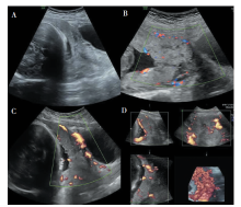
图1
超声图像 A:前置胎盘;B:胎盘增厚,肌层与胎盘基底部分界不清;C:三维能量图示胎盘后方丰富血流;D:三维血管成像示胎盘内血管走行分支紊乱。

表1
MRI及超声检查诊断PPP(n)
| 超声检查 | MRI检查 | 合计 | |
|---|---|---|---|
| PPP | 胎盘未见异常 | ||
| PPP | 122 | 2 | 124 |
| 胎盘未见异常 | 4 | 6 | 10 |
| 合计 | 126 | 8 | 134 |
表2
超声及MRI检查诊断PPP合并胎盘植入与临床病理诊断结果对照(n)
| 影像学检查 | 病理诊断 | |
|---|---|---|
| 未见胎盘植入(n=54) | 胎盘植入(n=80) | |
| 超声 | 61 | 73 |
| MRI | 59 | 75 |
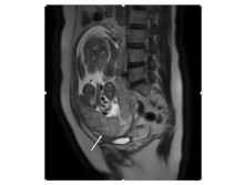
图2
MRI图像 T2WI序列矢状位图像示子宫肌层变薄,部分信号消失(箭头所示),局部结合带信号中断。

| [1] |
Chattopadhyay SK, Kharif H, Sherbeeni MM. Placenta praevia and accreta after previous caesarean section[J]. Eur J Obstet Gynecol Reprod Biol, 1993, 52(3):151-156.
pmid: 8163028 |
| [2] | Kazi S. Emergency peripartum hysterectomy: a great obstetric challenge[J]. Pak J Med Sci, 2018, 34(6):1567-1570. |
| [3] |
Berhan Y, Urgie T. A literature review of placenta accreta spectrum disorder: the place of expectant management in Ethiopian setup[J]. Ethiop J Health Sci, 2020, 30(2):277-292.
doi: 10.4314/ejhs.v30i2.16 pmid: 32165818 |
| [4] | 余琳, 胡可佳, 杨慧霞. 2008—2014年凶险性前置胎盘的回顾性临床研究[J]. 中华妇产科杂志, 2016, 51(3):169-173. |
| [5] |
Shih JC, Palacios Jaraquemada JM, et al. Role of three-dimensional power Doppler in the antenatal diagnosis of placenta accreta: comparison with gray-scale and color Doppler techniques[J]. Ultrasound Obstet Gynecol, 2009, 33(2):193-203.
doi: 10.1002/uog.6284 pmid: 19173239 |
| [6] |
Baughman WC, Corteville JE, Shah RR. Placenta accreta: spectrum of US and MR imaging findings[J]. Radiographics, 2008, 28(7):1905-1916.
doi: 10.1148/rg.287085060 URL |
| [7] |
Collins SL, Alemdar B, van Beekhuizen HJ, et al. Evidence-based guidelines for the management of abnormally invasive placenta: recommendations from the International Society for Abnormally Invasive Placenta[J]. Am J Obstet Gynecol, 2019, 220(6):511-526.
doi: S0002-9378(19)30433-8 pmid: 30849356 |
| [8] |
Angileri SA, Mailli L, Raspanti C, et al. Prophylactic occlusion balloon placement in internal iliac arteries for the prevention of postpartum haemorrhage due to morbidly adherent placenta: short term outcomes[J]. Radiol Med, 2017, 122(10):798-806.
doi: 10.1007/s11547-017-0777-z URL |
| [9] | 徐国华. 不同类型凶险性前置胎盘产妇的临床特点及剖宫产结局[J]. 中国妇幼保健, 2018, 33(9):1986-1988. |
| [10] | 刘彬, 高素娟, 张娜, 等. 核磁共振结合血清甲胎蛋白、肌酸激酶水平诊断凶险型前置胎盘合并胎盘植入的价值[J]. 中国妇幼保健, 2018, 33(6):1409-1412. |
| [11] | 杨彩群. 前置胎盘并发胎盘植入经腹彩色超声多普勒的诊断价值评价[J]. 中国妇幼健康研究, 2017, 28(S3):508-509. |
| [12] | 杨燕, 牛兆仪, 丁云川, 等. 妊娠晚期剖宫产瘢痕部位胎盘植入超声表现[J]. 中国超声医学杂志, 2015, 31(12):1111-1113. |
| [13] |
Yang JI, Lim YK, Kim HS, et al. Sonographic findings of placental lacunae and the prediction of adherent placenta in women with placenta previa totalis and prior cesarean section[J]. Ultrasound Obstet Gynecol, 2006, 28(2):178-182.
doi: 10.1002/uog.2797 URL |
| [14] | 郭吉敏, 曹满瑞, 刘小平, 等. MRI征象回归模型在植入型凶险性前置胎盘的应用[J]. 临床放射学杂志, 2018, 37(8):1325-1328. |
| [15] |
Dighe M. MR imaging of abnormal placentation[J]. Magn Reson Imaging Clin N Am, 2017, 25(3):601-610.
doi: 10.1016/j.mric.2017.03.002 URL |
| [16] | 宋慧玲, 蒋灵军, 蒋乐真, 等. 前置胎盘是否合并胎盘植入220例磁共振分析[J]. 中华医学杂志, 2018, 98(45):3692-3696. |
| [17] |
Maher MA, Abdelaziz A, Bazeed MF. Diagnostic accuracy of ultrasound and MRI in the prenatal diagnosis of placenta accreta[J]. Acta Obstet Gynecol Scand, 2013, 92(9):1017-1022.
doi: 10.1111/aogs.12187 URL |
| [18] | Goh WA, Zalud I. Placenta accreta: diagnosis, management and the molecular biology of the morbidly adherent placenta[J]. J Matern Fetal Neonatal Med, 2016, 29(11):1795-1800. |
| [19] |
Duzyj CM, Cooper A, Mhatre M, et al. Placenta accreta: aspectrum of predictable risk, diagnosis, and morbidity[J]. Am J Perinatol, 2019, 36(10):1031-1038.
doi: 10.1055/s-0038-1676111 URL |
| [1] | 黄娟, 朱晓雷, 李晓, 陈克敏, 严福华, 徐学勤. 血氧水平依赖磁共振成像评估早期慢性肾病肾缺氧的研究[J]. 诊断学理论与实践, 2022, 21(03): 385-389. |
| [2] | 朱乃懿, 姜奕歆, 柴丽, 柴维敏. 磁共振对超声阴性而乳腺X线检出BI-RADS4类以上钙化灶的诊断价值分析[J]. 诊断学理论与实践, 2021, 20(05): 439-444. |
| [3] | 张雪坤, 李彦, 严福华, 赵洪飞, 宋琦. 基于光梭成像的新型加速技术在颅脑MRI中的应用价值研究[J]. 诊断学理论与实践, 2021, 20(04): 378-383. |
| [4] | 吴萍, 冯炜炜. 剖宫产瘢痕妊娠的临床管理及继续妊娠的探究[J]. 诊断学理论与实践, 2020, 19(1): 95-99. |
| [5] | 吴霜, 解骞, 管雪妮, 张素芳, 高信芳, 梁宗辉. 磁共振体素内不相干运动扩散加权成像诊断活动期克罗恩病的价值及效能分析[J]. 诊断学理论与实践, 2020, 19(02): 157-161. |
| [6] | 曹烨, 刘晓晟, 葛晓乾, 周斌. 运用动态增强磁共振成像评估颈动脉粥样斑块稳定性的初步研究[J]. 诊断学理论与实践, 2019, 18(04): 436-441. |
| [7] | 朱晓雷, 陈璐, 陆文丽, 刘燕, 严福华, 王伟, 董治亚. 474例中枢性性早熟女童不同年龄段垂体MRI影像学异常比例分析[J]. 诊断学理论与实践, 2019, 18(03): 286-290. |
| [8] | 李云峰, 江泓, 李宁, 孙青芳. 核磁共振成像诊断三叉神经痛的价值分析与研究[J]. 诊断学理论与实践, 2018, 17(05): 562-565. |
| [9] | 许晶晶, 张敏鸣. 人工智能机器学习方法在阿尔茨海默病中的应用现状[J]. 诊断学理论与实践, 2018, 17(04): 466-470. |
| [10] | 赵华丽, 徐文鹏, 梁宗辉. 创伤性臂丛神经损伤的磁共振成像3D-FIESTA-C、IDEAL序列特征及诊断价值[J]. 诊断学理论与实践, 2018, 17(02): 197-201. |
| [11] | 黎鑫乐, 柴维敏, 朱樱. 乳腺乳头状肿瘤的影像学诊断进展[J]. 诊断学理论与实践, 2018, 17(02): 225-230. |
| [12] | 赵华丽, 席瑜玲, 韩春, 梁宗辉. 椎管内血管脂肪瘤的MRI表现[J]. 诊断学理论与实践, 2017, 16(06): 627-632. |
| [13] | 叶岚, 张欢, 钱朝霞. 磁共振在异位妊娠中的影像表现及临床价值[J]. 诊断学理论与实践, 2017, 16(06): 650-655. |
| [14] | 李旭东, 林晓珠, 房炜桓, 谢环环, 陈楠, 柴维敏, 严福华, 陈克敏. MRI图像纹理分析在胰腺神经内分泌肿瘤病理分级中的应用研究[J]. 诊断学理论与实践, 2017, 16(06): 601-606. |
| [15] | 苏明, 张继军, 凌华威, 张建, 丁蓓, 陆非, 刘燕. MRI检查在产后诊断胎盘植入中的价值[J]. 诊断学理论与实践, 2017, 16(05): 527-531. |
| 阅读次数 | ||||||
|
全文 |
|
|||||
|
摘要 |
|
|||||