诊断学理论与实践 ›› 2022, Vol. 21 ›› Issue (04): 476-481.doi: 10.16150/j.1671-2870.2022.04.010
车稳1, 柳蒋书1, 陈晓炎2( ), 王朝夫1, 袁菲1, 王璇1
收稿日期:2021-11-17
出版日期:2022-08-25
发布日期:2022-11-07
通讯作者:
陈晓炎
E-mail:cxy11832@rjh.com.cn
CHE Wen1, LIU Jiangshu1, CHEN Xiaoyan2(), WANG Chaofu1, YUAN Fei1, WANG Xuan1
Received:2021-11-17
Online:2022-08-25
Published:2022-11-07
Contact:
CHEN Xiaoyan
E-mail:cxy11832@rjh.com.cn
摘要:
目的:探讨肺混合性鳞状细胞和腺性乳头状瘤(pulmonary mixed squamous cell and glandular papilloma,PMSGP)的临床病理特征,并分析该病的冷冻切片病理诊断要点及误诊原因。方法:收集2019年至2021年上海交通大学医学院附属瑞金医院经术后石蜡切片病理检查确诊为PMSGP的病例2例,并结合文献分析其临床病理特点及冷冻切片病理检查误诊的原因。结果:2例患者分别为74岁的女性及66岁的男性,均为体检发现肺占位,其中1例伴有胸闷胸痛,影像学检查均考虑为恶性肿瘤,行肺叶切除术。术中冷冻切片病理检查结果,1例考虑为黏液腺癌,1例考虑为PMSGP,但不能排除黏液表皮样癌。术后石蜡切片病理检查显示,2例患者的肿瘤体积分别为3.0 cm×2.5 cm×2.5 cm和1.7 cm×1.7 cm×1.6 cm,肿瘤细胞排列呈乳头状,表面被覆腺上皮及鳞状上皮,乳头轴心有较多炎症细胞,鳞状上皮有轻-中度不典型性,核分裂象罕见,无坏死。免疫组织化学检查示,2例患者的肿瘤组织中,腺上皮细胞表达CK7和TTF-1,黏液柱状上皮表达MUC5AC,鳞状上皮及基底细胞表达CK5/6、P63和P40,Ki-67仅在基底层高表达。2例荧光原位杂交(fluorescence in situ hybridization,FISH)检测均未发现MAML2基因重排。2例患者分别随访9个月及24个月,均无复发或转移。复习相关文献报道的39例PMSGP患者,冷冻切片病理检查的肿瘤性质误诊率高达52.6%,误诊原因主要为PMSGP与黏液表皮样癌及黏液腺癌等恶性肿瘤存在形态学交叉,且被覆鳞状上皮的不典型性在冷冻切片病理诊断时容易被放大。结论:PMSGP为肺部少见的良性肿瘤,术中冷冻切片病理检查误诊率高。冷冻切片中,PMSGP肿瘤的形态特征包括境界清楚,呈乳头状结构,乳头被覆鳞状上皮及腺上皮,基底细胞沿纤维血管轴心栅栏状排列,细胞排列有层次感,无异型性。
中图分类号:
车稳, 柳蒋书, 陈晓炎, 王朝夫, 袁菲, 王璇. 肺混合性鳞状细胞和腺性乳头状瘤2例临床病理特征及冷冻切片病理诊断误诊分析[J]. 诊断学理论与实践, 2022, 21(04): 476-481.
CHE Wen, LIU Jiangshu, CHEN Xiaoyan, WANG Chaofu, YUAN Fei, WANG Xuan. Pulmonary mixed squamous cell and glandular papilloma clinicopathological characteristics of 2 cases and misdiagnosis analysis of frozen section[J]. Journal of Diagnostics Concepts & Practice, 2022, 21(04): 476-481.
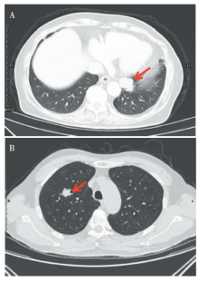
图1
胸部CT图像 A:病例1胸部增强CT示,左肺下叶后基底段软组织团块影,伴轻度均匀强化;B:病例2 胸部CT示,右肺上叶尖段类圆形病灶。
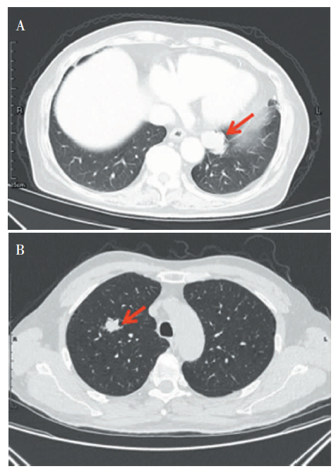
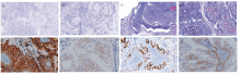
图2
病理图片 A:冷冻切片,低倍镜下肿瘤呈乳头状结构,乳头周围细胞层次多,部分细胞有不典型性;B:冷冻切片,肿瘤周围可见大片黏液湖,黏液湖内可见小簇状上皮细胞漂浮(箭头标示处);C:肿瘤位于段支气管腔内,呈乳头状结构,可见纤维血管轴心(×40);D:乳头被覆鳞状上皮和腺上皮(×100);E:腺上皮细胞CK7弥漫强阳性(EnVision法,×200);F:腺上皮细胞TTF-1阳性(EnVision法,×100);G 黏液柱状上皮强表达MUC5AC(EnVision法,×100);H. 鳞状细胞及基底样细胞表达P40(EnVision法,×100)。

表1
文献中PMSGP临床特征[例(n)]
| 文献 | 病例数 (男∶女) | 年龄(中位 年龄,岁) | 肿瘤位置 | 最大径(cm) | 分型 | 临床症状 | 随访 | ||
|---|---|---|---|---|---|---|---|---|---|
| 左肺 | 右肺 | 中央 | 外周 | ||||||
| Li等[ | 6(4∶2) | 39~67(53) | 1 | 5 | 1.8~4.2 | 1 | 5 | 腹胀、食欲不振1例;咳嗽咳痰1例;体检发现4例 | 随访12~52个月(平均31个月),其中1例术后11月胸部CT示双肺结节,考虑转移,其余均无复发转移 |
| Lin等[ | 5(2∶4) | 55~70(60) | 3 | 2 | 1.0~5.0 | 0 | 5 | 1例胸痛,1例胸痛及咳嗽,1例咳嗽及血痰,2例无症状 | 随访10~89个月(平均51.6个月),均无复发转移 |
| 汪小霞等[ | 5(0∶5) | 42~59(48) | 不详 | 不详 | 1.5~3.5 | 3 | 2 | 胸痛、胸闷、咳嗽咳白痰、痰中带血、发热 | 随访2~28个月,无复发转移 |
| Iijima等[ | 20(10∶10) | 34~84(63.5) | 10 | 10 | 0.9~3.0 | 8 | 12 | 不详 | 不详 |
表2
文献中PMSGP病例及冷冻切片病理诊断结果
| 文献 | 误诊率 | 冷冻病理诊断结果 |
|---|---|---|
| Lin等[ | 1/2 | 1例黏液表皮样癌,1例PMSGP |
| Iijima等[ | 6/12 | 2例腺癌,2例腺鳞癌,1例黏液表皮样癌,1例鳞状细胞癌,5例乳头状瘤,1例无恶性证据 |
| Yun等[ | 1/1 | 腺癌 |
| Miyoshi等[ | 1/1 | 黏液表皮样癌不能除外 |
| Feng等[ | 0/1 | 倾向良性病变 |
| 本文 | 1/2 | 1例黏液腺癌,1例考虑PMSGP,但不除外黏液表皮样癌 |
| [1] | Borczuk AC, Cooper WA, Dacic S, et al. WHO Classifica-tion of Tumours of Thoracic Tumours[M]. 5th ed, Lyon(France): International Agency for Research on Cancer, 2021:37-40. |
| [2] |
Flieder DB, Koss MN, Nicholson A, et al. Solitary pulmonary papillomas in adults: a clinicopathologic and in situ hybridization study of 14 cases combined with 27 cases in the literature[J]. Am J Surg Pathol, 1998, 22(11):1328-1342.
pmid: 9808125 |
| [3] |
Tryfon S, Dramba V, Zoglopitis F, et al. Solitary papillomas of the lower airways: epidemiological, clinical, and therapeutic data during a 22-year period and review of the literature[J]. J Thorac Oncol, 2012, 7(4):643-648.
doi: 10.1097/JTO.0b013e3182468d06 pmid: 22425912 |
| [4] | Noda N, Ebie Y, Matsumura M, et al. Comparison of detection specificity of nitrifying bacteria in biofilm using fluorescence in situ hybridization and in situ fluorescent antibody methods[J]. Water Sci Technol, 2003, 47(5):129-132. |
| [5] | Li F, He M, Li F, et al. Histologic characteristics and prognosis of lung mixed squamous cell and glandular papilloma: six case reports[J]. Int J Clin Exp Pathol, 2019, 12(9):3542-3548. |
| [6] |
Lin DL, Xing XM, Ran WW, et al. Pulmonary peripheral glandular papilloma and mixed squamous cell and glandular papilloma frequently harbour the BRAF V600E mutation[J]. Histopathology, 2020, 76(7):997-1004.
doi: 10.1111/his.14098 URL |
| [7] | 汪小霞, 李锐, 冯潇, 等. 肺混合性鳞状细胞和腺性乳头状瘤临床病理学分析[J]. 中华病理学杂志, 2019, 48(4):318-321. |
| Li R, Feng X, et al. Clinicopathological analysis of pulmonary mixed squamous cell and glandular papilloma[J]. Chin J Pathol, 2019, 48(4):318-321. | |
| [8] |
Iijima Y, Nakajima Y, Kinoshita H, et al. Mixed squamous cell and glandular papilloma of the lung: a case report and literature review in Japan[J]. Int J Surg Case Rep, 2020, 68:39-42.
doi: 10.1016/j.ijscr.2020.02.021 URL |
| [9] |
Yun JS, Kim DW, Choi YD, et al. Mixed squamous cell and glandular papilloma of the lung in a 64-year-old woman[J]. Korean J Thorac Cardiovasc Surg, 2014, 47(1):55-58.
doi: 10.5090/kjtcs.2014.47.1.55 pmid: 24570869 |
| [10] |
Miyoshi R, Menju T, Yoshizawa A, et al. Expression of p16 Ink4a in mixed squamous cell and glandular papilloma of the lung[J]. Pathol Int, 2017, 67(6):306-310.
doi: 10.1111/pin.12531 pmid: 28470939 |
| [11] | Feng AN, Wu HY, Zhou Q, et al. Solitary endobronchial papillomas with false impression of malignant transformation: report of two cases and review of the literature[J]. Int J Clin Exp Pathol, 2015, 8(7):8607-8612. |
| [12] | 刘荣美, 孟庆大. 肺混合性鳞状细胞和腺性乳头状瘤3例报道[J]. 诊断病理学杂志, 2018, 25(7):533-534. |
| Meng QD. Pulmonary mixed squamous cell and glandular papilloma: a report of three cases[J]. Chin J Diagn Pathol, 2018, 25(7):533-534. | |
| [13] |
Yabuki K, Matsuyama A, Obara K, et al. A unique case of a huge mixed squamous cell and glandular papilloma of non-endobronchial origin with a peripheral growth[J]. Respir Med Case Rep, 2018, 24:108-112.
doi: 10.1016/j.rmcr.2018.05.001 pmid: 29977775 |
| [14] | Huo Z, Wu H, Li J, et al. Primary pulmonary mucoepidermoid carcinoma: histopathological and moleculargenetic studies of 26 cases[J]. PLoS One, 2015, 10(11):e0143169. |
| [15] |
王征, 王恩华, 刘东戈. 肺原发性黏液性上皮源性肿瘤的病理诊断与鉴别诊断[J]. 中华肿瘤杂志, 2017, 39(1):1-6.
pmid: 28104025 |
|
Wang Z, Wang EH, Liu DG. Pathological diagnosis and differential diagnosis for primary pulmonary mucinous epi-thelial tumors[J]. Chin J Oncol, 2017, 39(1):1-6.
doi: 10.3760/cma.j.issn.0253-3766.2017.01.001 pmid: 28104025 |
|
| [16] |
Chang JC, Montecalvo J, Borsu L, et al. Bronchiolar adenoma: expansion of the concept of ciliated muconodular papillary tumors with proposal for revised terminology based on morphologic, immunophenotypic, and genomic analysis of 25 cases[J]. Am J Surg Pathol, 2018, 42(8):1010-1026.
doi: 10.1097/PAS.0000000000001086 pmid: 29846186 |
| [17] | Ishikawa Y. Ciliated muconodular papillary tumor of the peripheral lung: benign or malignant?[J]. Pathol Clin Med (Byouri-to-Rinsho), 2002,20,964-965. |
| [18] |
Kamata T, Sunami K, Yoshida A, et al. Frequent BRAF or EGFR mutations in ciliated muconodular papillary tumors of the lung[J]. J Thorac Oncol, 2016, 11(2):261-265.
doi: 10.1016/j.jtho.2015.10.021 pmid: 26718882 |
| [19] |
Liu L, Aesif SW, Kipp BR, et al. Ciliated muconodular papillary tumors of the lung can occur in western patients and show mutations in bRAF and AKT1[J]. Am J Surg Pathol, 2016, 40(12):1631-1636.
pmid: 27454941 |
| [20] |
Abe J, Ito S, Takahashi S, et al. Mixed squamous cell and glandular papilloma of the lung resembling early adenocarcinoma: A case report[J]. Ann Med Surg (Lond), 2016, 7:61-64.
doi: 10.1016/j.amsu.2016.03.025 pmid: 27141302 |
| [21] |
Sasaki E, Masago K, Fujita S, et al. AKT1 mutations in peripheral bronchiolar papilloma: glandular papilloma and mixed squamous cell and glandular papilloma is distinct from bronchiolar adenoma[J]. Am J Surg Pathol, 2021, 45(1):119-126.
doi: 10.1097/PAS.0000000000001573 URL |
| [22] | 陈晓炎, 杨晓群, 袁菲, 等. 肺纤毛黏液结节性乳头状肿瘤2例临床病理分析及文献复习[J]. 诊断学理论与实践, 2018, 17(5):575-580. |
| Yang XQ, Yuan F, et al. Pulmonary ciliated muconodular papillary tumor: clinical pathologic analysis of two cases and review of literature[J]. J Diagn Concepts & Pract, 2018, 17(5):575-580. | |
| [23] |
Popper HH, Wirnsberger G, Jüttner-Smolle FM, et al. The predictive value of human papilloma virus (HPV) typing in the prognosis of bronchial squamous cell papillomas[J]. Histopathology, 1992, 21(4):323-330.
pmid: 1328017 |
| [24] |
Huang YL, Chang YL, Chen KC, et al. Mixed squamous cell and glandular papilloma of the lung: a case report of a novel mutation in the BRAF gene and coexistent HPV infection, possible relationship to ciliated muconodular papillary tumor[J]. Pathol Int, 2019, 69(2):104-109.
doi: 10.1111/pin.12747 URL |
| [25] |
Inamura K, Kumasaka T, Furuta R, et al. Mixed squamous cell and glandular papilloma of the lung: a case study and literature review[J]. Pathol Int, 2011, 61(4):252-258.
doi: 10.1111/j.1440-1827.2011.02659.x pmid: 21418399 |
| [26] |
Tryfon S, Dramba V, Zoglopitis F, et al. Solitary papillomas of the lower airways: epidemiological, clinical, and therapeutic data during a 22-year period and review of the literature[J]. J Thorac Oncol, 2012, 7(4):643-648.
doi: 10.1097/JTO.0b013e3182468d06 pmid: 22425912 |
| [27] | Hayashi T, Tachibana S, Nakao K, et al. Solitary peripheral pulmonary squamous cell papilloma; diagnostic significance of 18F-fluorodeoxyglucose positron emission tomography findings[J]. Kyobu Geka, 2017, 70(4):309-312. |
| [1] | 李娟, 刘劲松, 李梅, 李殿炜, 朱弘. 细支气管腺瘤10例临床病理分析及文献复习[J]. 诊断学理论与实践, 2021, 20(05): 466-470. |
| [2] | 史冬梅, 王晓琳, 陈璐, 谢青. 曼氏裂头蚴病52例临床特点及误诊分析[J]. 诊断学理论与实践, 2020, 19(1): 37-43. |
| [3] | 王加临, 刘怡. 颈总动脉旁副甲状腺超声误诊1例分析[J]. 诊断学理论与实践, 2019, 18(06): 683-684. |
| [4] | 肖辅国, 潘自来. 浸润前病变的CT值变化在鉴别肺纯磨玻璃结节性质的价值探讨[J]. 诊断学理论与实践, 2019, 18(05): 521-525. |
| [5] | 赵伟伟, 王柳清, 张守成. 垂体催乳素瘤误诊为病毒性脑炎报告及文献复习[J]. 诊断学理论与实践, 2018, 17(06): 723-725. |
| [6] | 王荟, 刘坤, 陈佳, 房振, 高扬, 吴丽莉. 甲状腺髓样癌细针穿刺被误诊为甲状腺乳头状癌1例报道[J]. 诊断学理论与实践, 2018, 17(03): 337-340. |
| [7] | 张姗姗, 杨玲. 肺栓塞误诊为肺炎的原因分析[J]. 诊断学理论与实践, 2017, 16(05): 549-552. |
| [8] | 吴云林, 毛峻岭, 吴巍, 黄天生, 陆敏, 陈晓敏, 罗方秀, 陆亭伟, 袁晓琴, 李佑, 项明,. 2013年上海嘉定地区胃镜筛查胃癌漏诊的临床研究[J]. 诊断学理论与实践, 2014, 13(04): 383-387. |
| [9] | 郑叶, 童海涛, 曾东, 杨月香, 冯艳玲, 卢洪洲,. 艾滋病合并眼、胰腺和脾结核误诊为肿瘤3例临床与病理分析[J]. 诊断学理论与实践, 2014, 13(02): 187-192. |
| [10] | 郭海英, 包婺平, 周新,. 隐源性机化性肺炎误诊社区获得性肺炎24例并文献复习[J]. 诊断学理论与实践, 2013, 12(02): 166-169. |
| [11] | 邹静, 刘斌, 陈雪华, 李宁丽, 王利, 查琼芳,. 肺癌患者外周血调节性T细胞检测的临床意义[J]. 诊断学理论与实践, 2010, 9(03): 260-263. |
| [12] | 赵华丽, 柴维敏, 汪登斌, 李蔚, 陈克敏,. 乳腺良、恶性病变的磁共振成像误诊分析[J]. 诊断学理论与实践, 2009, 8(03): 309-313. |
| [13] | 沈军, 蔡颖, 徐潜, 曹兴午, 鄢盛恺,. 恶性疟原虫感染1例的误诊分析[J]. 诊断学理论与实践, 2009, 8(03): 338-340. |
| [14] | 陆晓兰, 蒋小平, 周世红, 叶玉梅, 樊华,. 反复误诊为子宫黏膜下血管纤维瘤的内膜间质肉瘤[J]. 诊断学理论与实践, 2009, 8(02): 200-201. |
| [15] | 杨道华, 沈铭昌,. 肺嗜酸细胞类癌1例[J]. 诊断学理论与实践, 2006, 5(03): 261-. |
| 阅读次数 | ||||||
|
全文 |
|
|||||
|
摘要 |
|
|||||