Journal of Diagnostics Concepts & Practice ›› 2019, Vol. 18 ›› Issue (2): 133-138.doi: 10.16150/j.1671-2870.2019.02.003
• Original articles • Previous Articles Next Articles
CHEN Jie1, HU Jin2, YANG Kang1, FU Yi1
Received:2019-02-20
Online:2019-04-25
Published:2019-04-25
CLC Number:
CHEN Jie, HU Jin, YANG Kang, FU Yi. Analysis of risk factors and prognosis of cerebral hemorrhage patients accompanied by cortical superficial siderosis[J]. Journal of Diagnostics Concepts & Practice, 2019, 18(2): 133-138.
| 参数 | 深部出血组 (n=185) | 脑叶出血组 (n=56) | P 值 |
|---|---|---|---|
| 基础情况 年龄(岁) | 61±12 | 66±12 | 0.007 |
| 性别(男性) | 121 (65.4%) | 39 (69.6%) | 0.556 |
| 吸烟 | 62 (33.5%) | 18 (32.1%) | 0.839 |
| 饮酒 | 46 (24.9%) | 14 (25.0%) | 1.000 |
| 糖尿病 | 26 (14.1%) | 11 (19.6%) | 0.309 |
| 高血压病 | 175 (94.6%) | 43 (76.8%) | <0.001 |
| 口服药物 | |||
| 降压药物 | 160 (86.5%) | 39 (69.6%) | 0.004 |
| 抗血小板药物 | 21 (11.4%) | 7 (12.5%) | 0.814 |
| 实验室数据 | |||
| 胆固醇(mmol/L) | 4.7±1.1 | 4.7±1.1 | 0.777 |
| 三酰甘油(mmol/L) | 1.9±1.5 | 1.5±0.9 | 0.011 |
| 高密度脂蛋白(mmol/L) | 1.2±0.8 | 1.3±0.3 | 0.797 |
| 低密度脂蛋白(mmol/L) | 3.0±0.9 | 3.0±0.8 | 0.618 |
| 空腹血糖(mmol/L) | 6.1±2.3 | 6.1±1.7 | 0.890 |
| 尿素氮(mmol/L) | 5.5±3.3 | 5.3±1.5 | 0.607 |
| 肌酐(μmol/L) | 69.0 (56.0~81.0) | 69.0 (56.5~83.7) | 0.407 |
| 同型半胱氨酸(mmol/L) | 16.7±10.6 | 14.6±6.5 | 0.205 |
| 入院血压(mmHg) | |||
| 收缩压 | 155.6±22.2 | 145.0±21.9 | 0.002 |
| 舒张压 | 89.6±12.7 | 82.4±15.5 | 0.001 |
| 平均动脉压 | 111.0±16.5 | 101.7±21.0 | 0.001 |
| 影像学数据 | |||
| 脑血肿量(mL) | 4.0 (1.2~9.7) | 9.9 (3.3~20) | <0.001 |
| cSS | 12 (6.5%) | 13 (23.2%) | <0.001 |
| 白质高信号 | 147 (79.5%) | 45 (80.4%) | 0.884 |
| 无症状性脑梗死灶 | 55 (29.7%) | 22 (39.3%) | 0.179 |
| CMB存在 | 123 (66.5%) | 33 (58.9%) | 0.300 |
| CMBs(个) | 2 (0~5) | 1 (0~5) | 0.246 |
| 3个月mRS评分>2分 | 29 (15.8%) | 7 (12.5%) | 0.550 |
| 参数 | cSS (-)(n=26) | cSS (+)(n=16) | P值 |
|---|---|---|---|
| 基础情况 年龄(岁) | 71±10 | 68±10 | 0.293 |
| 性别(男性) | 18 (69.2%) | 14 (87.5%) | 0.177 |
| 吸烟 | 11 (42.3%) | 5 (31.3%) | 0.474 |
| 饮酒 | 7 (26.9%) | 5 (31.3%) | 1.000 |
| 糖尿病 | 4 (15.4%) | 4 (25.0%) | 0.441 |
| 高血压病 | 17 (65.4%) | 12 (75.0%) | 0.756 |
| 口服药物 | |||
| 降压药物 | 17 (65.4%) | 12 (75.0%) | 0.756 |
| 抗血小板药物 | 2 (7.7%) | 4 (25.0%) | 0.270 |
| 入院血压(mmHg) | |||
| 收缩压 | 150.8±23.4 | 146.4±15.2 | 0.508 |
| 舒张压 | 81.0±11.6 | 86.5±11.2 | 0.135 |
| 平均动脉压 | 104.3±13.4 | 93.5±37.9 | 0.288 |
| 影像学数据 | |||
| 脑血肿量(mL) | 11.0 (2.2~20.0) | 14.4 (1.0~24.8) | 0.742 |
| 白质高信号 | 26 (100%) | 16 (100%) | |
| 无症状性脑梗死 | 11 (42.3%) | 7 (43.8%) | 0.927 |
| CMB存在 | 15 (57.7%) | 16 (100%) | 0.008 |
| CMBs数目(个) | 1 (0~3) | 2 (1~7) | 0.053 |
| 3个月mRS评分>2分 | 4 (15.4%) | 4 (25.0%) | 0.714 |
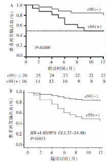
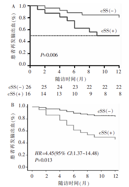
| 1年内死亡或再发 | 4 (15.4%) | 9 (56.3%) | 0.015 |
| [1] | 中华医学会神经病学分会, 中华医学会神经病学分会脑血管病学组. 中国脑出血诊治指南(2014)[J]. 中华神经科杂志, 2015, 48(6):435-444. |
| [2] | 中华医学会神经外科学分会, 中国医师协会急诊医师分会, 国家卫生和计划生育委员会脑卒中筛查与防治工程委员会. 自发性脑出血诊断治疗中国多学科专家共识[J]. 中华神经外科杂志, 2015, 31(12): 1189-1194. |
| [3] | Caetano A, Ladeira F, Barbosa R, et al. Cerebral amyloid angiopathy - The modified Boston criteria in clinical practice[J]. J Neurol Sci, 2018, 384:55-57. |
| [4] | DeSimone CV, Graff-Radford J, El-Harasis MA, et al. Cerebral Amyloid Angiopathy: Diagnosis, Clinical Implications, and Management Strategies in Atrial Fibrillation[J]. J Am Coll Cardiol, 2017, 70(9):1173-1182. |
| [5] | Charidimou A, Gang Q, Werring DJ. Sporadic cerebral amyloid angiopathy revisited: recent insights into pathophysiology and clinical spectrum[J]. J Neurol Neurosurg Psychiatry, 2012, 83(2):124-137. |
| [6] | 冯玉兰, 蒋爱华, 颜静, 等. 脑血肿体积评估3种方法的比较研究[J]. 实用临床医药杂志, 2014, 18(11):26-30,36. |
| [7] | Greenberg SM, Vernooij MW, Cordonnier C, et al. Cerebral microbleeds: a guide to detection and interpretation[J]. Lancet Neurol, 2009, 8(2):165-174. |
| [8] | Cheng CY, Cheng HM, Chen SP, et al. White matter hyperintensities in migraine: Clinical significance and central pulsatile hemodynamic correlates[J]. Cephalalgia, 2018, 38(7):1225-1236. |
| [9] | Gupta A, Giambrone AE, Gialdini G, et al. Silent Brain Infarction and Risk of Future Stroke: A Systematic Review and Meta-Analysis[J]. Stroke, 2016, 47(3):719-725. |
| [10] | Charidimou A, Linn J, Vernooij MW, et al. Cortical superficial siderosis: detection and clinical significance in cerebral amyloid angiopathy and related conditions[J]. Brain, 2015, 138(Pt 8):2126-2139. |
| [11] | Broderick JP, Adeoye O, Elm J. Evolution of the Modified Rankin Scale and Its Use in Future Stroke Trials[J]. Stroke, 2017, 48(7):2007-2012. |
| [12] | Yaghi S, Rostanski SK, Boehme AK, et al. Imaging Parameters and Recurrent Cerebrovascular Events in Patients With Minor Stroke or Transient Ischemic Attack[J]. JAMA Neurol, 2016, 73(5):572-578. |
| [13] | 时景璞, 滕卫禹, 王海龙, 等. 高血压高发区人群脑卒中患病现状的流行病学调查[J]. 中国卫生统计, 2006, 23(1):47-49. |
| [14] | Pantoni L. Cerebral small vessel disease: from pathoge-nesis and clinical characteristics to therapeutic challenges[J]. Lancet Neurol, 2010, 9(7):689-701. |
| [15] | Cordonnier C, Al-Shahi Salman R, Wardlaw J. Spontaneous brain microbleeds: systematic review, subgroup analyses and standards for study design and reporting[J]. Brain, 2007, 130(Pt 8):1988-2003. |
| [16] | Greenberg SM, Nandigam RN, Delgado P, et al. Microbleeds versus macrobleeds: evidence for distinct entities[J]. Stroke, 2009, 40(7):2382-2386. |
| [17] | Linn J, Herms J, Dichgans M, et al. Subarachnoid hemosiderosis and superficial cortical hemosiderosis in cerebral amyloid angiopathy[J]. Am J Neuroradiol, 2008, 29(1):184-186. |
| [18] | Linn J, Michl S, Katja B, et al. Cortical vein thrombosis: the diagnostic value of different imaging modalities[J]. Neuroradiology, 2010, 52(10):899-911. |
| [19] | Kumar S, Goddeau RP, Jr, Selim MH, et al. Atraumatic convexal subarachnoid hemorrhage: clinical presentation, imaging patterns, and etiologies[J]. Neurology, 2010, 74(11):893-899. |
| [20] | Na HK, Park JH, Kim JH, et al. Cortical superficial siderosis: a marker of vascular amyloid in patients with cognitive impairment[J]. Neurology, 2015, 84(8):849-855. |
| [21] | Samarasekera N, Smith C, Al-Shahi Salman R. The association between cerebral amyloid angiopathy and intrace-rebral haemorrhage: systematic review and meta-analysis[J]. J Neurol Neurosurg Psychiatry, 2012, 83(3):275-281. |
| [22] | Hemphill JC 3rd, Greenberg SM, Anderson CS, et al. Guidelines for the Management of Spontaneous Intrace-rebral Hemorrhage: A Guideline for Healthcare Professionals From the American Heart Association/American Stroke Association[J]. Stroke, 2015, 46(7):2032-2060. |
| [23] | Wermer MJH, Greenberg SM. The growing clinical spectrum of cerebral amyloid angiopathy[J]. Curr Opin Neurol, 2018, 31(1):28-35. |
| [24] | Wieberdink RG, Poels MM, Vernooij MW, et al. Serum lipid levels and the risk of intracerebral hemorrhage: the Rotterdam Study[J]. Arterioscler Thromb Vasc Biol, 2011, 31(12):2982-2989. |
| [25] | Phuah CL, Raffeld MR, Ayres AM, et al. Subacute decline in serum lipids precedes the occurrence of primary intracerebral hemorrhage[J]. Neurology, 2016, 86(22):2034-2041. |
| [26] | Shoamanesh A, Martinez-Ramirez S, Oliveira-Filho J, et al. Interrelationship of superficial siderosis and microbleeds in cerebral amyloid angiopathy[J]. Neurology, 2014, 83(20):1838-1843. |
| [1] | HUANG Juan, ZHU Xiaolei, LI Xiao, CHEN Kemin, YAN Fuhua, XU Xueqin. Study on blood oxygen level-dependent magnetic resonance imaging for the assessment of early renal hypoxia in chronic kidney disease [J]. Journal of Diagnostics Concepts & Practice, 2022, 21(03): 385-389. |
| [2] | ZHU Naiyi, JIANG Yixin, CHAI Li, CHAI Weimin. Diagnostic values of magnetic resonance imaging in mammography detected BI-RADS≥4 category calcifications with negative ultrasound results [J]. Journal of Diagnostics Concepts & Practice, 2021, 20(05): 439-444. |
| [3] | ZHANG Xuekun, LI Yan, YAN Fuhua, ZHAO Hongfei, SONG Qi. Application value of new accelerating technology based on constellation shuttling imaging in brain MRI [J]. Journal of Diagnostics Concepts & Practice, 2021, 20(04): 378-383. |
| [4] | SUN Tiantian, YE Baoying, YANG Yu, NIU Jianmei. Color Doppler ultrasound and magnetic resonance imaging in prenatal diagnosis of pernicious placenta previa and pernicious placenta previa with placenta accreta: clinic value and analysis of missed diagnosis [J]. Journal of Diagnostics Concepts & Practice, 2021, 20(02): 173-177. |
| [5] | CAO Juntao, HU Ming, QIAN Pingkang, TU Jianchun, ZHANG Huan, SHEN Junkang. Application value of 3.0T MRI 3D-MERGE sequence in evaluating the degree of supraspinatus tendon injury [J]. Journal of Diagnostics Concepts & Practice, 2021, 20(01): 77-81. |
| [6] | WU Shuang, XIE Qian, GUAN Xueni, ZHANG Sufang, GAO Xinfang, LIANG Zonghui. Perfomence of MRI intravoxel incoherent motion diffusion weighted imaging parameters in diagnosing active Crohn's disease [J]. Journal of Diagnostics Concepts & Practice, 2020, 19(02): 157-161. |
| [7] | GU Xiaohong, SUN Aimin, WANG Qian, ZHU Ming, ZHONG Yumin. The three-dimensional balanced steady state free precession magnetic resonance imaging sequence in diagnosis of anomalous origin of the coronary artery from the pulmonary artery in children [J]. Journal of Diagnostics Concepts & Practice, 2020, 19(02): 145-150. |
| [8] | CAO Ye, LIU Xiaosheng, GE Xiaoqian, ZHOU Bin. Preliminary study on dynamic contrast-enhanced MRI in identifying vulnerability of carotid atherosclerotic plaques [J]. Journal of Diagnostics Concepts & Practice, 2019, 18(04): 436-441. |
| [9] | LI Xinyue, TAN Lin, CHAI Weimin. Value of MRI combined with DWI in differential diagnosis of benign papillary lesions with malignant papillary lesions of breast [J]. Journal of Diagnostics Concepts & Practice, 2019, 18(03): 301-306. |
| [10] | ZHU Xiaolei, CHEN Lu, LU Wenli, LIU Yan, YAN Fuhua, WANG Wei, DONG Zhiya. Radiological findings on pituitary MRI in central precocious puberty [J]. Journal of Diagnostics Concepts & Practice, 2019, 18(03): 286-290. |
| [11] | LI Yunfeng, JIANG Hong, LI Ning, SUN Qingfang. Analysis and study on value of MRI in diagnosis of trigeminal neuralgia [J]. Journal of Diagnostics Concepts & Practice, 2018, 17(05): 562-565. |
| [12] | ZHAO Hua-li, XU Wenpeng, LIANG Zonghui. The features and diagnostic value of 3D-FIESTA-C and IDEAL sequences for brachial plexus injury [J]. Journal of Diagnostics Concepts & Practice, 2018, 17(02): 197-201. |
| [13] | YE Lan, ZHANG Huan, QIAN Zhaoxia. Features of magnetic resonance imaging and its value in clinicaldiagnosis of ectopic pregnancy [J]. Journal of Diagnostics Concepts & Practice, 2017, 16(06): 650-655. |
| [14] | ZHAO Huali, XI Yuling, HAN Chun, LIANG Zonghui. Analysis of MRI findings of spinal angiolipoma [J]. Journal of Diagnostics Concepts & Practice, 2017, 16(06): 627-632. |
| [15] | SU Ming, ZHANG Jijun, LING Huawei, ZHANG Jian, DING Bei, LU Fei, LIU Yan. Diagnostic value of MRI for postpartum placenta implantation [J]. Journal of Diagnostics Concepts & Practice, 2017, 16(05): 527-531. |
| Viewed | ||||||
|
Full text |
|
|||||
|
Abstract |
|
|||||