Journal of Diagnostics Concepts & Practice ›› 2019, Vol. 18 ›› Issue (03): 301-306.doi: 10.16150/j.1671-2870.2019.03.011
• Original articles • Previous Articles Next Articles
LI Xinyue, TAN Lin( ), CHAI Weimin
Received:2019-06-10
Online:2019-06-25
Published:2019-06-25
Contact:
TAN Lin
E-mail:tl10927@rjh.com.cn
CLC Number:
LI Xinyue, TAN Lin, CHAI Weimin. Value of MRI combined with DWI in differential diagnosis of benign papillary lesions with malignant papillary lesions of breast[J]. Journal of Diagnostics Concepts & Practice, 2019, 18(03): 301-306.
| 组别 | 良性 | 恶性 | P值 |
|---|---|---|---|
| 平均年龄(岁) | 49.4 | 60.4 | |
| 病灶数(个) | 106 | 40 | |
| 肿块样强化 | 74(69.8%) | 16(40.0%) | <0.001 |
| 非肿块强化 | 32(30.2%) | 24(60.0%) |
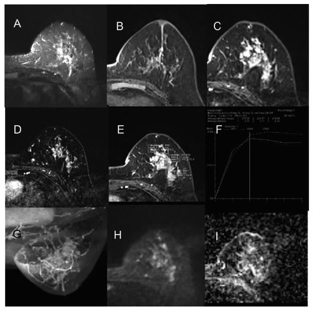
| 肿块 | 良性(n=74) | 恶性(n=16) | P值 |
|---|---|---|---|
| 形态 | 0.003 | ||
| 圆形、卵圆形 | 28(37.8) | 0 | |
| 不规则 | 46(62.2) | 16(100) | |
| 边缘 | <0.001 | ||
| 光滑 | 40(54.1) | 0 | |
| 不光滑 | 31(41.9) | 7(43.7) | |
| 毛刺 | 3(4.0) | 9(56.3) | |
| 直径 | <0.001 | ||
| <1 cm | 44(59.5) | 0 | |
| 1~5 cm | 30(40.5) | 16(100) | |
| 位置 | 0.904 | ||
| 乳晕后区 | 22(29.7) | 5(31.2) | |
| 非乳晕后区 | 52(70.3) | 11(68.8) | |
| 强化方式 | <0.001 | ||
| 均匀 | 35(47.3) | 0 | |
| 不均匀 | 25(33.8) | 12(75.0) | |
| 环形 | 14(18.9) | 4(25.0) |

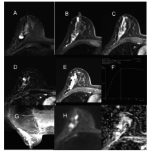
| 非肿块型病灶 | 良性(n=32) | 恶性(n=24) | P值 |
|---|---|---|---|
| 分布 | 0.036 | ||
| 局灶 | 12(37.5) | 3(12.5) | |
| 节段 | 18(56.3) | 15(62.5) | |
| 区域或弥漫 | 2(6.3) | 6(25.0) | |
| 伴囊性灶 | 0.120 | ||
| 伴 | 5(15.6) | 8(33.3) | |
| 不伴 | 27(84.4) | 16(66.7) | |
| 强化方式 | 0.516 | ||
| 均匀 | 3(9.4) | 2(9.3) | |
| 不均匀 | 9(28.1) | 10(41.7) | |
| 集簇 | 19(59.4) | 10(41.7) | |
| 成簇小环状 | 1(3.1) | 2(8.3) |


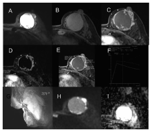
| 肿块型+非肿块型 | 良性(n=106) | 恶性(n=40) | P值 |
|---|---|---|---|
| TIC | 0.004 | ||
| 上升型 | 30(28.3) | 2(5.0) | |
| 平台型 | 29(27.4) | 10(25.0) | |
| 流出型 | 47(44.3) | 28(70.0) | |
| 导管扩张 | 0.148 | ||
| 扩张 | 66(62.3) | 30(75.0) | |
| 不扩张 | 40(37.7) | 10(25.0) | |
| 实性占比 | 0.028 | ||
| <25% | 2(1.9) | 4(10.0) | |
| >25% | 104(98.1) | 36(90.0) |

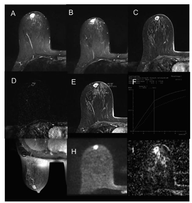
| 参数 | 良性 | 恶性 | P值 |
|---|---|---|---|
| 早期强化率平均值(%) | 214 | 255 | 0.049 |
| 峰值强化率平均值(%) | 346 | 348 | 0.843 |
| ADC值平均值(×10-3 mm2/s) | 1.13 | 0.95 | <0.05 |
| [1] | 杨文涛. 乳腺乳头状病变的诊断与鉴别诊断[J]. 诊断病理学杂志, 2008, 15(4):257-260. |
| [2] |
Boufelli G, Giannotti MA, Ruiz CA, et al. Papillomas of the breast: factors associated with underestimation[J]. Eur J Cancer Prev, 2018, 27(4):310-314.
doi: 10.1097/CEJ.0000000000000343 URL |
| [3] |
Kestelman FP, Gomes CF, Fontes FB, et al. Imaging findings of papillary breast lesions: a pictorial review[J]. Clin Radiol, 2014, 69(4):436-441.
doi: 10.1016/j.crad.2013.11.020 pmid: 24457016 |
| [4] | 阮玫, 赵亚娥, 汪登斌, 等. 乳腺导管内乳头状瘤的乳腺专用磁共振成像表现及其诊断价值[J]. 放射学实践, 2013, 28(3):341-345. |
| [5] | Sarica O, Uluc F, Tasmali D. Magnetic resonance imagi-ng features of papillary breast lesions[J]. Send to Eur J Radiol, 2014, 83(3):524-530. |
| [6] |
Kurz KD, Roy S, Saleh A, Diallo-Danebrock R, et al. MRI features of intraductal papilloma of the breast: sheep in wolf's clothing?[J]. Acta Radiol, 2011, 52(3):264-272.
doi: 10.1258/ar.2011.100434 URL |
| [7] |
Bhattarai N, Kanemaki Y, Kurihara Y, et al. Intraductal papilloma: features on MR ductography using a microscopic coil[J]. Am J Roentgenol, 2006, 186(1):44-47.
pmid: 16357375 |
| [8] |
Zhu Y, Zhang S, Liu P, et al. Solitary intraductal papillomas of the breast: MRI features and differentiation from small invasive ductal carcinomas[J]. Am J Roentgenol, 2012, 199(4):936-942.
doi: 10.2214/AJR.12.8507 URL |
| [9] |
Yildiz S, Toprak H, Ersoy YE, et al. Contribution of diffusion-weighted imaging to dynamic contrast-enhanced MRI in the characterization of papillary breast lesions[J]. Breast J, 2018, 24(2):176-179.
doi: 10.1111/tbj.12861 URL |
| [10] |
Guo S, Wang Y, Rohr J, et al. Solid papillary carcinoma of the breast: A special entity needs to be distinguished from conventional invasive carcinoma avoiding over-treatment[J]. Breast, 2016, 26:67-72.
doi: 10.1016/j.breast.2015.12.015 URL |
| [11] |
Linda A, Zuiani C, Girometti R, et al. Unusual malignant tumors of the breast: MRI features and pathologic correlation[J]. Eur J Radiol, 2010, 75(2):178-184.
doi: 10.1016/j.ejrad.2009.04.038 pmid: 19446418 |
| [12] |
Brookes MJ, Bourke AG. Radiological appearances of papillary breast lesions[J]. Clin Radiol, 2008, 63(11):1265-1273.
doi: 10.1016/j.crad.2008.02.012 pmid: 18929044 |
| [13] |
Kuhl C. The current status of breast MR imaging. Part I. Choice of technique, image interpretation, diagnostic accuracy, and transfer to clinical practice[J]. Radiology, 2007, 244(2):356-378.
doi: 10.1148/radiol.2442051620 URL |
| [14] |
Kul S, Cansu A, Alhan E, et al. Contribution of diffusion-weighted imaging to dynamic contrast-enhanced MRI in the characterization of breast tumors[J]. Am J Roentgenol, 2011, 196(1):210-217.
doi: 10.2214/AJR.10.4258 URL |
| [1] | HUANG Juan, ZHU Xiaolei, LI Xiao, CHEN Kemin, YAN Fuhua, XU Xueqin. Study on blood oxygen level-dependent magnetic resonance imaging for the assessment of early renal hypoxia in chronic kidney disease [J]. Journal of Diagnostics Concepts & Practice, 2022, 21(03): 385-389. |
| [2] | YUE Jingjing, SONG Qi, JIANG Xufeng, WANG Li, ZHAO Weili, YAN Fuhua. Comparison of magnetic resonance whole body diffusion weighted imaging with FS-T2WI and FDG-PET/CT for initial staging and detection of lesion in newly diagnosed lymphoma [J]. Journal of Diagnostics Concepts & Practice, 2021, 20(06): 540-546. |
| [3] | ZHU Naiyi, JIANG Yixin, CHAI Li, CHAI Weimin. Diagnostic values of magnetic resonance imaging in mammography detected BI-RADS≥4 category calcifications with negative ultrasound results [J]. Journal of Diagnostics Concepts & Practice, 2021, 20(05): 439-444. |
| [4] | ZHANG Xuekun, LI Yan, YAN Fuhua, ZHAO Hongfei, SONG Qi. Application value of new accelerating technology based on constellation shuttling imaging in brain MRI [J]. Journal of Diagnostics Concepts & Practice, 2021, 20(04): 378-383. |
| [5] | SUN Tiantian, YE Baoying, YANG Yu, NIU Jianmei. Color Doppler ultrasound and magnetic resonance imaging in prenatal diagnosis of pernicious placenta previa and pernicious placenta previa with placenta accreta: clinic value and analysis of missed diagnosis [J]. Journal of Diagnostics Concepts & Practice, 2021, 20(02): 173-177. |
| [6] | CAO Juntao, HU Ming, QIAN Pingkang, TU Jianchun, ZHANG Huan, SHEN Junkang. Application value of 3.0T MRI 3D-MERGE sequence in evaluating the degree of supraspinatus tendon injury [J]. Journal of Diagnostics Concepts & Practice, 2021, 20(01): 77-81. |
| [7] | WU Shuang, XIE Qian, GUAN Xueni, ZHANG Sufang, GAO Xinfang, LIANG Zonghui. Perfomence of MRI intravoxel incoherent motion diffusion weighted imaging parameters in diagnosing active Crohn's disease [J]. Journal of Diagnostics Concepts & Practice, 2020, 19(02): 157-161. |
| [8] | GU Xiaohong, SUN Aimin, WANG Qian, ZHU Ming, ZHONG Yumin. The three-dimensional balanced steady state free precession magnetic resonance imaging sequence in diagnosis of anomalous origin of the coronary artery from the pulmonary artery in children [J]. Journal of Diagnostics Concepts & Practice, 2020, 19(02): 145-150. |
| [9] | CHEN Jie, HU Jin, YANG Kang, FU Yi. Analysis of risk factors and prognosis of cerebral hemorrhage patients accompanied by cortical superficial siderosis [J]. Journal of Diagnostics Concepts & Practice, 2019, 18(2): 133-138. |
| [10] | CAO Ye, LIU Xiaosheng, GE Xiaoqian, ZHOU Bin. Preliminary study on dynamic contrast-enhanced MRI in identifying vulnerability of carotid atherosclerotic plaques [J]. Journal of Diagnostics Concepts & Practice, 2019, 18(04): 436-441. |
| [11] | ZHU Xiaolei, CHEN Lu, LU Wenli, LIU Yan, YAN Fuhua, WANG Wei, DONG Zhiya. Radiological findings on pituitary MRI in central precocious puberty [J]. Journal of Diagnostics Concepts & Practice, 2019, 18(03): 286-290. |
| [12] | LI Yunfeng, JIANG Hong, LI Ning, SUN Qingfang. Analysis and study on value of MRI in diagnosis of trigeminal neuralgia [J]. Journal of Diagnostics Concepts & Practice, 2018, 17(05): 562-565. |
| [13] | ZHAO Hua-li, XU Wenpeng, LIANG Zonghui. The features and diagnostic value of 3D-FIESTA-C and IDEAL sequences for brachial plexus injury [J]. Journal of Diagnostics Concepts & Practice, 2018, 17(02): 197-201. |
| [14] | YE Lan, ZHANG Huan, QIAN Zhaoxia. Features of magnetic resonance imaging and its value in clinicaldiagnosis of ectopic pregnancy [J]. Journal of Diagnostics Concepts & Practice, 2017, 16(06): 650-655. |
| [15] | ZHAO Huali, XI Yuling, HAN Chun, LIANG Zonghui. Analysis of MRI findings of spinal angiolipoma [J]. Journal of Diagnostics Concepts & Practice, 2017, 16(06): 627-632. |
| Viewed | ||||||
|
Full text |
|
|||||
|
Abstract |
|
|||||