Journal of Diagnostics Concepts & Practice ›› 2019, Vol. 18 ›› Issue (2): 139-143.doi: 10.16150/j.1671-2870.2019.02.004
• Original articles • Previous Articles Next Articles
LI Weiwei1, ZHAN Weiwei2, ZHOU Wei2( ), TAO Lingling1, WANG Yi1, FAN Jinfang1, FEI Yuanxin1, KUANG Lijun1, XU Wenying1
Received:2019-01-01
Online:2019-04-25
Published:2019-04-25
Contact:
ZHOU Wei
E-mail:zw11468@126.com
CLC Number:
LI Weiwei, ZHAN Weiwei, ZHOU Wei, TAO Lingling, WANG Yi, FAN Jinfang, FEI Yuanxin, KUANG Lijun, XU Wenying. The diagnostic value of smart three-dimensional superb microvascular imaging in detecting blood flow distribution pattern of breast lesion[J]. Journal of Diagnostics Concepts & Practice, 2019, 18(2): 139-143.
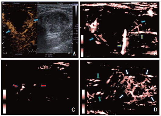
| CEUS对比剂灌注 | Smart 3D SMI | ||||
|---|---|---|---|---|---|
| 点线型(n=11) | 分枝型(n=32) | 球形型(n=58) | 扩散型(n=20) | 混合型(n=24) | |
| 灌注与否 | |||||
| 有 | 11 | 32 | 58 | 20 | 24 |
| 无 | 0 | 0 | 0 | 0 | 0 |
| 粗大血流 | |||||
| 有 | 6 (55%) | 24 (75%) | 42(72%) | 16(80%) | 19(79%) |
| 无 | 5 | 8 | 16 | 4 | 5 |
| 分支血流 | |||||
| 有 | 2 | 7 | 19 | 8 | 10 |
| 无 | 9(82%) | 25 (78%) | 39 (67%) | 12 (60%) | 14(58%) |
| 强度 | |||||
| 强 | 5 (45%) | 27 (84%)(p1) | 50 (86%) (P2) (P3) | 15 (75%) | 17 (71%) |
| 中或弱 | 6 | 5 | 8 | 5 | 7 |
| 均匀与否 | |||||
| 是 | 4 (36%) | 15 (47%)(p4) | 36 (62%) (P5) (P6) | 6 (30%) | 8 (33%) |
| 否 | 7 | 17 | 22 | 14 | 16 |
| 位于肿块 | |||||
| 内部 | 11 | 29 | 54 | 0 | 0 |
| 周边扩散状 | 0 | 0 | 0 | 14 | 0 |
| 内及周边 | 0 | 3 | 4 | 6 | 24 |

| [1] | 孙可欣, 李贺, 孙可欣, 等. 2014年中国恶性肿瘤发病和死亡分析[J]. 中华肿瘤杂志, 2018, 40(1):5. |
| [2] | Wang M, Feng HL, Liu YQ, et al. Angiogenesis Research in Mouse Mammary Cancer Based on Contrast-enhanced Ultrasonography: Exploratory Study[J]. Acad Radiol, 2018, 25(7):889-897. |
| [3] | Holmgren L, O'Reilly MS, Folkman J. Dormancy of micrometastases: balanced proliferation and apoptosis in the presence of angiogenesis suppression[J]. Nat Med, 1995, 1(2):149-153. |
| [4] | Yongfeng Z, Ping Z, Wengang L, et al. Application of a Novel Microvascular Imaging Technique in Breast Lesion Evaluation[J]. Ultrasound Med Biol, 2016, 42(9):2097-2105. |
| [5] | Park AY, Seo BK. Up-to-date Doppler techniques for breast tumor vascularity: superb microvascular imaging and contrast-enhanced ultrasound[J]. Ultrasonography, 2018, 37(2):98-106. |
| [6] | Adler DD, Carson PL, Rubin JM, et al. Doppler ultrasound color flow imaging in the study of breast cancer: preliminary findings[J]. Ultrasound Med Biol, 1990, 16(6):553-559. |
| [7] | Li Q, Hu M, Chen Z, et al. Meta-Analysis: Contrast-Enhanced Ultrasound Versus Conventional Ultrasound for Differentiation of Benign and Malignant Breast Lesions[J]. Ultrasound Med Biol, 2018, 44(5):919-929. |
| [8] | Wan CF, Liu XS, Wang L, et al. Quantitative contrast-enhanced ultrasound evaluation of pathological complete response in patients with locally advanced breast cancer receiving neoadjuvant chemotherapy[J]. Eur J Radiol, 2018, 103:118-123. |
| [9] | Wang XY, Kang LK, Lan CY. Contrast-enhanced ultrasonography in diagnosis of benign and malignant breast lesions[J]. Eur J Gynaecol Oncol, 2014, 35(4):415-420. |
| [10] | Machado P, Segal S, Lyshchik A, et al. A Novel Microvascular Flow Technique: Initial Results in Thyroids[J]. Ultrasound Q, 2016, 32(1):67-74. |
| [11] | Mao Y, Mu J, Zhao J, et al. The value of superb microvascular imaging in differentiating benign renal mass from malignant renal tumor: a retrospective study[J]. Br J Radiol, 2018, 91(1082):20170601. |
| [12] | Karaca L, Oral A, Kantarci M, et al. Comparison of the superb microvascular imaging technique and the color Doppler techniques for evaluating children's testicular blood flow[J]. Eur Rev Med Pharmacol Sci, 2016, 20(10):1947-1953. |
| [13] | He MN, Lv K, Jiang YX, et al. Application of superb microvascular imaging in focal liver lesions[J]. World J Gastroenterol, 2017, 23(43):7765-7775. |
| [14] | Ma Y, Li G, Li J, et al. The Diagnostic Value of Superb Microvascular Imaging (SMI) in Detecting Blood Flow Signals of Breast Lesions: A Preliminary Study Compari-ng SMI to Color Doppler Flow Imaging[J]. Medicine (Baltimore), 2015, 94(36):e1502. |
| [15] | 朱慧, 徐卫平, 陈红燕, 等. 超声造影联合超微血管显像对乳腺癌微血管的评价[J]. 中国临床医学影像杂志, 2016, 27(6):390-392. |
| [16] | 陈欣, 肖晓云, 吴欢, 等. 微细血流成像技术在乳腺肿瘤鉴别中的应用[J]. 中国超声医学杂志, 2016, 32(5):407-410. |
| [1] | DIAO Xuehong, SHEN Yan, CHEN Lin, ZHAN Jia, FANG Liang, CAI Jianfei, CHEN Yue. Application of superb microvascular imaging technology in diagnosing rheumatoid arthritis in the clinical remission stage [J]. Journal of Diagnostics Concepts & Practice, 2022, 21(05): 575-580. |
| [2] | WANG Zhiqian, LI Min, YÜ Yifei, ZHOU Jianqiao. Ultrasonographic characteristics of testicular adrenal residue tumor in patients with congenital adrenal hyperplasia due to 21-hydroxylase deficiency [J]. Journal of Diagnostics Concepts & Practice, 2022, 21(05): 588-591. |
| [3] | GU Xuan, LIU Jun. Ultrasound screening to identify solid pseudopapillary tumours of the pancreas from pancreatic ductal adenocarcinoma [J]. Journal of Diagnostics Concepts & Practice, 2022, 21(04): 504-508. |
| [4] | HE Xin, CHEN Hui, FENG Weiwei. Research progress on the application of machine learning in assisted ultrasound diagnosis of adnexal masses [J]. Journal of Diagnostics Concepts & Practice, 2022, 21(04): 541-546. |
| [5] | DU Yanran, JIAO Jing, REN Yunyun, ZHOU Jianqiao. Application of ultrasound-based radiomics technology in the evaluation of fetal lung maturity [J]. Journal of Diagnostics Concepts & Practice, 2022, 21(03): 326-330. |
| [6] | CAO Yunyun, WANG Guanjie, ZENG Min, WANG Haifeng, NIU Jianmei, ZHOU Leiping. Prediction value of first trimester ultrasound parameters for pregnancy outcome [J]. Journal of Diagnostics Concepts & Practice, 2021, 20(05): 445-449. |
| [7] | YANG Tian, JI Xiang, NIU Jianmei, KONG Xiaoxiao, LV Mingli. Application of 2D-ultrasonography in prenatal assessment of fetal thymus development [J]. Journal of Diagnostics Concepts & Practice, 2021, 20(05): 471-474. |
| [8] | ZHAO Ran, ZHAN Weiwei, LIU Jun. Value of three-dimensional ultrasonic monitoring testicular volume for predicting spermatogenic function in patients with idiopathic hypogonadotropic hypogonadism [J]. Journal of Diagnostics Concepts & Practice, 2021, 20(03): 279-283. |
| [9] | SUN Tiantian, YE Baoying, YANG Yu, NIU Jianmei. Color Doppler ultrasound and magnetic resonance imaging in prenatal diagnosis of pernicious placenta previa and pernicious placenta previa with placenta accreta: clinic value and analysis of missed diagnosis [J]. Journal of Diagnostics Concepts & Practice, 2021, 20(02): 173-177. |
| [10] | LI Weiwei, WU Ying, ZHOU Wei, ZHAN Weiwei, ZHOU Qinghua, TAO Lingling, YANG Yanwen. The diagnostic value of smart three-dimensional superb microvascular imaging in differentiating benign and malignant breast lesions of BI-RADS 4 [J]. Journal of Diagnostics Concepts & Practice, 2020, 19(06): 583-587. |
| [11] | WANG Xing, WANG Ronghui, ZHANG Guiping, DONG Yijie, ZHOU Wei, ZHAN Weiwei. Diagnostic accuracy of ultrasound-guided fine needle aspiration biopsy for different thyroid carcinoma in 10 388 thyroid nodules: a ten-year study [J]. Journal of Diagnostics Concepts & Practice, 2020, 19(04): 359-363. |
| [12] | HONG Guiping, CHEN Xiaoyan, ZHOU Jianping, CHEN Wei, XIANG Yi, ZHOU Min, LI Qingyun. Diagnostic value of endobronchial ultrasound-guided transbronchial needle aspiration in elderly patients with hilar and mediastinal lesions and analysis of missed diagnosis [J]. Journal of Diagnostics Concepts & Practice, 2020, 19(04): 397-401. |
| [13] | FEI Yuanxin, KUANG Lijun, LU Caifeng, LI Weiwei, ZHOU Wei, ZHOU Qinghua. Sonographic findings of thyroid tuberculosis: Report of a case and review of literature [J]. Journal of Diagnostics Concepts & Practice, 2020, 19(03): 269-273. |
| [14] | CHEN Xiaoying, ZHENG Jie. Establishment of pretibial myxedema severity and activity rating scale and validation with ultrasonic and laboratory measurements [J]. Journal of Diagnostics Concepts & Practice, 2020, 19(03): 243-247. |
| [15] | XU Shangyan, JIA Xiaohong, NI Xiaofeng, ZHAN Weiwei. Interobserver variability in sonographic evaluation of thyroid nodules with ACR-TIRADS and RJ-TIRADS [J]. Journal of Diagnostics Concepts & Practice, 2019, 18(2): 149-154. |
| Viewed | ||||||
|
Full text |
|
|||||
|
Abstract |
|
|||||