Journal of Diagnostics Concepts & Practice ›› 2025, Vol. 24 ›› Issue (02): 155-162.doi: 10.16150/j.1671-2870.2025.02.006
Previous Articles Next Articles
CHANG Rui, LI Jiqiang, YANG Yanzhao, CHAI Weimin, YAN Fuhua, DONG Haipeng.( )
)
Received:2025-01-07
Accepted:2025-03-08
Online:2025-04-25
Published:2025-07-11
Contact:
DONG Haipeng.
E-mail:dhp40427@rjh.com.cn
CLC Number:
CHANG Rui, LI Jiqiang, YANG Yanzhao, CHAI Weimin, YAN Fuhua, DONG Haipeng.. Evaluation value of single-phase images from photon-counting CT-based low-dose pancreatic dynamic volume perfusion scanning for pancreatic cancer imaging[J]. Journal of Diagnostics Concepts & Practice, 2025, 24(02): 155-162.
Table 1
Acquisition and reconstruction parameters for photon-counting CT spancreatic dynamic volume perfusion imaging
| Acquisition parameters Parameter | |
|---|---|
| Scan mode | PCCT(NAEOTOM Alpha) |
| Tube voltage (kVp) | 90 |
| Image quality (IQ) level | 80 |
| Tube current | Care Dose 4D |
| Spiral pitch | 1.0 |
| Rotation time(s) | 0.25 |
| Collimation(mm) | 144 × 0.4 |
| Total acquisition time(s) | 47(began at 7) |
| Z-axis coverage *(cm) | 16 - 25 |
| Number of scan cycles † | 17 - 23 |
| Contrast Volume(mL) | 40 |
| Iodine concentration(mg/mL) | 400 |
| Flow rate(mL/s) | 4 |
| Reconstruction Parameters | |
| Quantum iterative reconstruction | QIR 4 |
| Reconstruction filter kernel | Br 40 |
| Slice thickness and increment (mm) | 1 / 1 |
| Image reconstruction | 55 keV / 70 keV / T3D |
Figure 1
Individual phase image extraction from photon-counting CT volume perfusion CT scanningA: After automatic motion correction and 4D noise reduction, regions of interest (ROIs) were placed in the normal pancreatic parenchyma and portal vein; B: Time-attenuation curves were generated. The first scan cycle from the perfusion dataset served as the noncontrast images, whereas the highest enhancement points of the pancreatic parenchyma and portal vein were manually selected as individual phase images for routine diagnostic interpretation and morphological assessment. C–E: Case of PDAC located in the pancreatic tail. Individual phase images reconstructed from 55 keV VMI, noncontrast phase (C), pancreatic parenchymal phase (D), and portal venous phase (E).
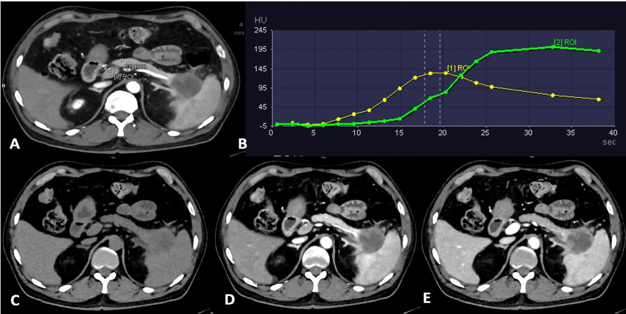
Table 2
Characteristics and Radiation Dose of the Study Population(N=55)
| Parameter | Value |
|---|---|
| Sex | Male 34,Female 21 |
| Age(y) | 66.4 ± 6.6 (49-78) |
| Weight(kg) | 62.5 ± 10.2 (40-83) |
| Height(m) | 1.66 ± 0.90 (1.45-1.86) |
| BMI(kg/m2) | 22.7 ± 3.2 (17.6-32.9) |
| Z-axis range(cm) | 18.7 ± 2.2 (16-25) |
| No. of scan cycles | 20 ± 2.0 (17-23) |
| CTDIvol(mGy) | 58.7 ± 14.6 (28.3-95.4) |
| DLP(mGy · cm) | 1199.4 ± 328.8 (472.5-1 943.1) |
| ED(mSv) | 18.0 ± 4.9 (7.1-29.2) |
Figure 2
Subjective image quality scores of individual phase image from photon-counting CT volume perfusion scanning
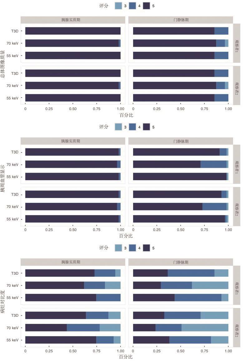
Table 3
Objective evaluation of image quality of individual phase image from photon-counting CT volume perfusion scanning
| Item | T3D | 70 keV | 55 keV | P * | P† | P ‡ | P§ |
|---|---|---|---|---|---|---|---|
| Pancreatic parenchymal phase | |||||||
| Image noise (HU) | 8.2 ± 2.1 | 7.1 ± 1.9 | 8.3 ± 2.1 | < 0.001 | < 0.001 | 0.599 | < 0.001 |
| SNR parenchyma | 16.3 ± 5.9 | 16.7 ± 5.0 | 18.8 ± 6.4 | < 0.001 | 0.002 | < 0.001 | < 0.001 |
| SNR vessel | 35.9 ± 15.9 | 36.1 ± 12.5 | 46.8 ± 19.8 | < 0.001 | < 0.001 | < 0.001 | < 0.001 |
| SNR lesion | 7.2 ± 1.8 | 7.5 ± 2.1 | 7.6 ± 2.0 | 0.036 | 0.062 | 0.003 | 0.505 |
| CNR vessel | 27.0 ± 17.7 | 25.7 ± 16.2 | 36.9 ± 23.0 | < 0.001 | < 0.001 | < 0.001 | < 0.001 |
| CNR lesion | 9.1 ± 3.7 | 8.0 ± 3.2 | 11.1 ± 4.4 | < 0.001 | < 0.001 | < 0.001 | < 0.001 |
| Portal venous phase | |||||||
| Image noise (HU) | 8.3 ± 2.2 | 7.3 ± 1.8 | 8.4 ± 2.1 | < 0.001 | < 0.001 | 0.683 | < 0.001 |
| SNR parenchyma | 12.4 ± 3.2 | 11.8 ± 3.2 | 13.3 ± 3.6 | < 0.001 | 0.033 | < 0.001 | < 0.001 |
| SNR vessel | 20.9 ± 6.3 | 18.5 ± 5.0 | 23.3 ± 7.5 | < 0.001 | < 0.001 | < 0.001 | < 0.001 |
| SNR lesion | 6.5 ± 2.0 | 6.7 ± 1.9 | 6.9 ± 2.2 | 0.003 | 0.009 | < 0.001 | 0.231 |
| CNR vessel | 10.6 ± 4.4 | 9.0 ± 3.7 | 13.5 ± 5.7 | < 0.001 | < 0.001 | < 0.001 | < 0.001 |
| CNR lesion | 5.7 ± 3.0 | 4.9 ± 2.7 | 6.3 ± 3.0 | < 0.001 | < 0.001 | < 0.001 | < 0.001 |
| [1] | LI H O, SUN C, XU Z D, et al. B. Low-dose whole organ CT perfusion of the pancreas: preliminary study[J]. Abdom Imaging,2014,39(1):40-7. |
| [2] |
O'MALLEY R B, SOLOFF E V, COVELER A L, et al. Feasibility of wide detector CT perfusion imaging performed during routine staging and restaging of pancreatic ductal adenocarcinoma[J]. Abdom Radiol (NY),2021,46(5):1992-2002.
doi: 10.1007/s00261-020-02786-y pmid: 33079256 |
| [3] |
O'MALLEY R B, COX D, SOLOFF E V, et al. CT perfusion as a potential biomarker for pancreatic ductal adenocarcinoma during routine staging and restaging[J]. Abdom Radiol (NY),2022,47(11):3770-3781.
doi: 10.1007/s00261-022-03638-7 pmid: 35972550 |
| [4] | PERIK T H, VAN GENUGTEN E A J, AARNTZEN E H J G, et al. Quantitative CT perfusion imaging in patients with pancreatic cancer: a systematic review[J]. Abdom Radiol (NY),2022,47(9):3101-3117. |
| [5] | SKORNITZKE S, VATS N, MAYER P, et al. Pancreatic CT perfusion: quantitative meta-analysis of disease discrimination, protocol development, and effect of CT parameters[J]. Insights Imaging,2023,14(1):132. |
| [6] |
HAMDY A, ICHIKAWA Y, TOYOMASU Y, et al. Perfusion CT to assess response to neoadjuvant chemotherapy and radiation therapy in pancreatic ductal adenocarcinoma: initial experience[J]. Radiology,2019,292(3):628-635.
doi: 10.1148/radiol.2019182561 pmid: 31287389 |
| [7] | WANG X, HENZLER T, GAWLITZA J, et al. Image qua-lity of mean temporal arterial and mean temporal portal venous phase images calculated from low dose dynamic volume perfusion CT datasets in patients with hepatocellular carcinoma and pancreatic cancer[J]. Eur J Radiol,2016,85(11):2104-2110. |
| [8] |
WILLEMINK M J, PERSSON M, POURMORTEZA A, et al. Photon-counting CT: technical principles and clinical prospects[J]. Radiology,2018,289:293-312
doi: 10.1148/radiol.2018172656 pmid: 30179101 |
| [9] |
FLOHR T, SCHMIDT B. Technical basics and clinical benefits of photon-counting CT[J]. Invest Radiol,2023,58:441-450
doi: 10.1097/RLI.0000000000000980 pmid: 37185302 |
| [10] |
LI J, CHEN X Y, XU K, et al. Detection of insulinoma: one-stop pancreatic perfusion CT with calculated mean temporal images can replace the combination of bi-phasic plus perfusion scan[J]. Eur Radiol,2020,30(8):4164-4174.
doi: 10.1007/s00330-020-06657-4 pmid: 32189051 |
| [11] | MICHALSKI C W, ERKAN M, HUSER N, et al. Resection of primary pancreatic cancer and liver metastasis: a systematic review[J]. Dig Surg,2008,25(6):473–480 |
| [12] | KLEIN F, PUHL G, GUCKELBERGER O, et al. The impact of simultaneous liver resection for occult liver metastases of pancreatic adenocarcinoma[J]. Gastroenterol Res Pract,2012(6):939-950 |
| [13] | LENG S, BRUESEWITZ M, TAO S, et al. Photon-coun-ting detector CT: system design and clinical applications of an emerging technology[J]. Radiographics,2019,39(3):729-743. |
| [14] |
KIM J, MABUD T, HUANG C, et al. Inter-reader agreement of pancreatic adenocarcinoma resectability assessment with photon counting versus energy integrating detector CT[J]. Abdom Radiol (NY),2024,49(9):3149-3157.
doi: 10.1007/s00261-024-04298-5 pmid: 38630314 |
| [15] | 谢环环, 林晓珠, 王晴柔, 等. CT能谱成像在胰腺癌病灶显示中的应用价值[J].实用放射学杂志,2017,33(5):750-753. |
| XIE H H, LIN X Z, WANG Q R, et al. Value of CT spectral imaging in demonstration of pancreatic ductal adenocarcinoma[J]. J Pract Radiol, 2017, 33(5):750-753. | |
| [16] | BEER L, TOEPKER M, BA-SALAMAH A, et al. Objective and subjective comparison of virtual monoenergetic vs. polychromatic images in patients with pancreatic ductal adenocarcinoma[J]. Eur Radiol,2019,29(7):3617-3625. |
| [17] | 杨琰昭, 常蕊, 王晴柔, 等. 双层探测器光谱CT虚拟单能量图像在胰腺导管腺癌术前评估中的优化研究[J]. 上海交通大学学报:医学版, 2022, 42(9):1323-1328. |
| YANG Y Z, CHANG R, WANG Q R, et al. Optimized study of virtual monoenergetic images derived from a dual-layer spectral detector CT in the preoperative evalua-tion of pancreatic ductal adenocarcinoma[J]. J Shanghai Jiaotong Univ(Med Sci),2022,(9):1323-1328 | |
| [18] | DECKER J A, BECKER J, HARTING M, et al. Optimal conspicuity of pancreatic ductal adenocarcinoma in virtual monochromatic imaging reconstructions on a photon-counting detector CT: comparison to conventional MDCT[J]. Abdom Radiol (NY),2024,49(1):103-116. |
| [1] | LÜ Haiying, LU Yong, HE Naying. Clinical applications of photon-counting CT in neuroimaging [J]. Journal of Diagnostics Concepts & Practice, 2025, 24(02): 212-219. |
| [2] | CAI Xinxin, DENG Rong, XU Xinxin, XU Zhihan, CHANG Rui, DONG Haipeng, LIN Huimin, YAN Fuhua. Study on consistency between liver fat fraction quantification based on photon-counting CT and MRI proton density fat fraction [J]. Journal of Diagnostics Concepts & Practice, 2025, 24(02): 146-154. |
| [3] | HUANG Ruikun, YANG Yanzhao, CHAI Weimin. Advances in application of photon-counting CT for pancreatic imaging [J]. Journal of Diagnostics Concepts & Practice, 2025, 24(02): 111-117. |
| [4] | LÜ Xiaoyu, FENG Weiming, ZHOU Huiyun, LI Jiqiang, DONG Haipeng, HUANG Juan. Feasibility of reducing scan time based on deep learning reconstruction in magnetic resonance imaging: a phantom study [J]. Journal of Diagnostics Concepts & Practice, 2024, 23(02): 131-138. |
| [5] | FAN Jing, YANG Wenjie, WANG Mengzhen, LU Wei, SHI Xiaomeng, ZHU Hong. The application of deep learning algorithm reconstruction in low tube voltage coronary CT angiography [J]. Journal of Diagnostics Concepts & Practice, 2022, 21(03): 374-379. |
| [6] | ZHANG Xuekun, LI Yan, YAN Fuhua, ZHAO Hongfei, SONG Qi. Application value of new accelerating technology based on constellation shuttling imaging in brain MRI [J]. Journal of Diagnostics Concepts & Practice, 2021, 20(04): 378-383. |
| [7] | WANG Tao, FU Meng, XIAO Ruijie, DONG Haipeng, LI Ruokun, YAN Fuhua. Application of Multivane XD technique in liver T2-weighted imaging [J]. Journal of Diagnostics Concepts & Practice, 2016, 15(05): 521-524. |
| [8] | . [J]. Journal of Diagnostics Concepts & Practice, 2011, 10(06): 531-534. |
| [9] | . [J]. Journal of Diagnostics Concepts & Practice, 2011, 10(02): 158-161. |
| [10] | . [J]. Journal of Diagnostics Concepts & Practice, 2010, 9(02): 141-145. |
| [11] | . [J]. Journal of Diagnostics Concepts & Practice, 2010, 9(02): 155-160. |
| [12] | . [J]. Journal of Diagnostics Concepts & Practice, 2008, 7(06): 637-640. |
| [13] | . [J]. Journal of Diagnostics Concepts & Practice, 2008, 7(01): 42-46. |
| [14] | . [J]. Journal of Diagnostics Concepts & Practice, 2004, 3(03): 34-37. |
| Viewed | ||||||
|
Full text |
|
|||||
|
Abstract |
|
|||||