Journal of Diagnostics Concepts & Practice ›› 2024, Vol. 23 ›› Issue (05): 537-541.doi: 10.16150/j.1671-2870.2024.05.011
• Case report • Previous Articles Next Articles
WANG Yurong1,2, WANG Yuanyuan1,2, WENG Haiyan1,2( )
)
Received:2024-04-14
Accepted:2024-08-06
Online:2024-10-25
Published:2025-02-25
Contact:
WENG Haiyan
E-mail:Whaiyan1166@163.com
CLC Number:
WANG Yurong, WANG Yuanyuan, WENG Haiyan. Clinical and pathological analysis of gastrointestinal leiomyosarcoma:Report of three cases[J]. Journal of Diagnostics Concepts & Practice, 2024, 23(05): 537-541.
Table 1
The clinical and pathological data of 3 cases of leiomyosarcoma
| 例号 | 性别 | 年龄(岁) | 肿块大小(cm) | 部位 | 临床表现 | 治疗 | 随访(月) |
|---|---|---|---|---|---|---|---|
| 1 | 女 | 62 | 4.5×4.0×4.0 | 胃底部 | 食欲下降伴体重减轻 | 手术切除 | 复发(30) |
| 2 | 男 | 68 | 4.5×3.5×2.5 | 小肠 | 持续性左侧肢体麻木 | 手术切除 | 无复发(24) |
| 3 | 女 | 58 | 7.5×5.0×4.5 | 胃体小弯侧 | 上腹部饱胀 | 手术切除 | 无复发(11) |
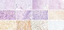
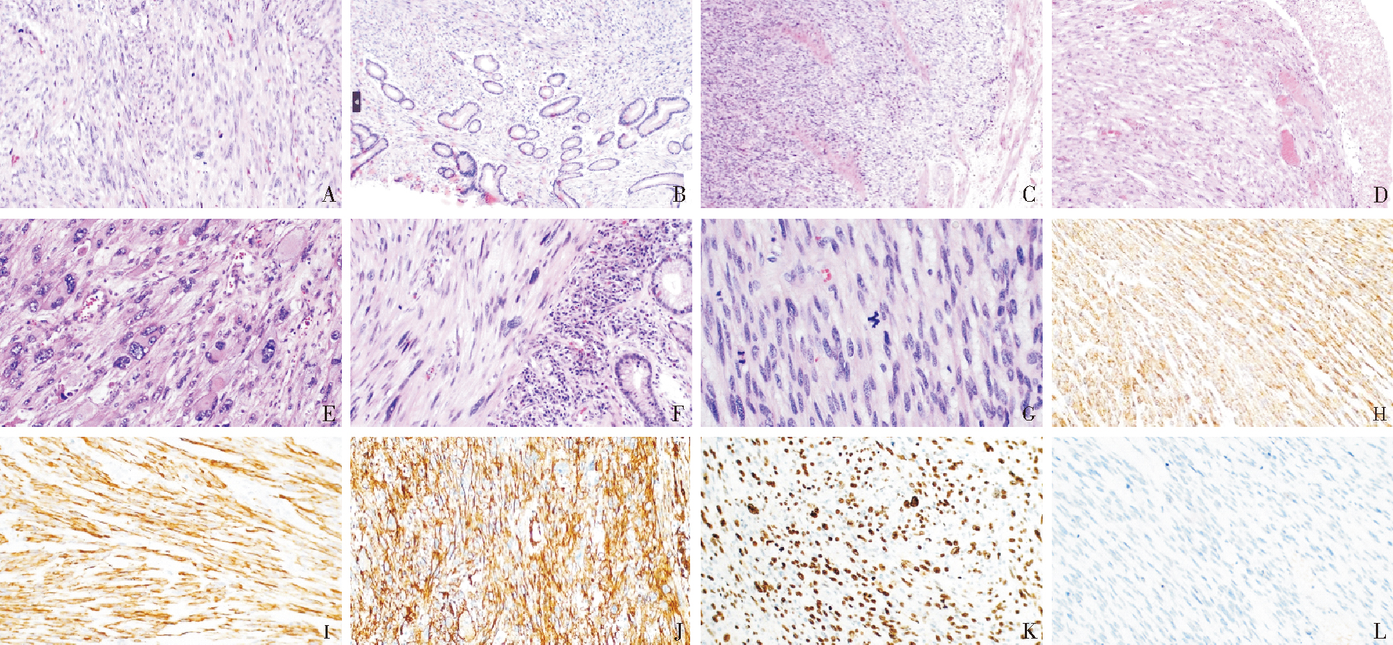
Figure 1
Gastrointestinal leiomyosarcoma A. The tumor cells are spindle shaped and can be seen arranged in bundles or interweaves HE, ×200; B. Tumor cells invading the mucosal lamina propria HE, ×100; C. Tumor cells interpenetrate between the intrinsic muscle layer HE, ×100; D. Bleeding, necrosis, and granulation tissue hyperplasia can be seen on the mucosal surface HE, ×100; E. Tumor giant cell HE, ×200; F. Singular nucleus HE, ×200; G. mitotic figure HE, ×400; H. SMA diffuse positive expression (EnVision two-step method, ×200); I. Desmin diffuse positive expression (EnVision two-step method, ×200); J. Caldesmon diffuse positive expression (EnVision two-step method, ×200); K. Ki-67 proliferation index expression EnVision two-step method, ×200; L. CD117 negative expression EnVision two-step method, ×200
| [1] | SERRANO C, GEORGE S. Leiomyosarcoma[J]. Hematol Oncol Clin North Am, 2013, 27(5):957-974. |
| [2] |
GEORGE S, SERRANO C, HENSLEY M L, et al. Soft tissue and uterine leiomyosarcoma[J]. J Clin Oncol, 2018, 36(2):144-150.
doi: 10.1200/JCO.2017.75.9845 pmid: 29220301 |
| [3] | KANG W Z, XUE L Y, TIAN Y T. Leiomyosarcoma of the stomach: A case report[J]. World J Clin Cases, 2019, 7 (21):3575-3582. |
| [4] |
AGGARWAL G, SHARMA S, ZHENG M, et al. Primary leiomyosarcomas of the gastrointestinal tract in the post-gastrointestinal stromal tumor era[J]. Ann Diagn Pathol, 2012, 16(6):532-540.
doi: 10.1016/j.anndiagpath.2012.07.005 pmid: 22917807 |
| [5] |
TAKAGI T, SAITO S, YOKOTA S, et al. Laparoscopic and endoscopic cooperative surgery for leiomyosarcoma of the stomach: a case report with a review of the literature[J]. Surg Case Rep, 2021, 7(1):146.
doi: 10.1186/s40792-021-01218-3 pmid: 34143361 |
| [6] | BANANZADEH A, MOKHTARI M, SOHOOLI M, et al. Two cases of primary leiomyosarcoma of sigmoid colon treated with laparoscopic surgery: A case report and a review of literature[J]. Int J Surg Case Rep, 2021, 87:106420. |
| [7] | WANG T, ZREIK R, LENG B. The landscape of primary gastric leiomyosarcoma in texas population: analysis of texas cancer registry data[J]. Cureus, 2023, 15(11):e49403. |
| [8] | ANNICCHIARICO A, MONTALI F, BALDINU M, et al. Leiomyosarcoma of the rectum: A systematic review of recent literature[J]. J Surg Oncol, 2024, 129(2):365-380. |
| [9] |
ARTS R, BOSSCHA K, RANSCHAERT E, et al. Small bowel leiomyosarcoma: a case report and literature review[J]. Turk J Gastroenterol, 2012, 23(4):381-384.
pmid: 22965511 |
| [10] |
MIETTINEN M, LASOTA J. Gastrointestinal stromal tumors: review on morphology, molecular pathology, prognosis, and differential diagnosis[J]. Arch Pathol Lab Med, 2006, 130(10):1466-1478.
doi: 10.5858/2006-130-1466-GSTROM pmid: 17090188 |
| [11] | BASU I, LEMONAS P. Leiomyosarcoma of the rectum following pelvic irradiation: a difficult histological diagnosis[J]. Ann R Coll Surg Engl, 2012, 94(1):e44-e45 |
| [12] | CRYSTAL J S, KORDERAS K, SCHWARTZBERG D, et al. Primary leiomyosarcoma of the colon: a report of two cases, review of the literature, and association with immunosuppression for IBD and rheumatoid arthritis[J]. Case Rep Surg, 2018, 2018:6824643. |
| [13] |
MARUZZO M, BRUNELLO A, DIMINUTTO A, et al. Long-term response to first-line trabectedin in an elderly female patient with a metastatic leiomyosarcoma unfit for anthracycline[J]. Anticancer Drugs, 2016, 27(3):264-267.
doi: 10.1097/CAD.0000000000000326 pmid: 26629769 |
| [14] | COCO C, RIZZO G, MANNO A, et al. Surgical treatment of small bowel neoplasms[J]. Eur Rev Med Pharmacol Sci, 2010, 14(4):327-333. |
| [15] | ÖZTEKIN M, YILMAZ B, AĞAGÜNDÜZ D, et al. Overview of helicobacter pylori infection: clinical features, treatment, and nutritional aspects. diseases[J]. 2021, 9(4):66. |
| [16] | GARCIA-ORTEGA D Y, REYES-GARCIA N, MARTINEZ-SAID H, et al. Radiation-induced leiomyosarcoma of the rectum after cervical cancer treatment[J]. Rev Gastroenterol Mex (Engl Ed), 2018, 83(4):465-467. |
| [17] |
ANDERSON N D, BABICHEV Y, FULIGNI F, et al. Lineage-defined leiomyosarcoma subtypes emerge years before diagnosis and determine patient survival[J]. Nat Commun, 2021, 12(1):4496.
doi: 10.1038/s41467-021-24677-6 pmid: 34301934 |
| [18] |
Cancer Genome Atlas Research Network. Electronic address: elizabeth.demicco@sinaihealthsystem.ca; Cancer Genome Atlas Research Network. Comprehensive and Integrated Genomic Characterization of Adult Soft Tissue Sarcomas[J]. Cell, 2017, 171(4):950-965.e28.
doi: S0092-8674(17)31203-5 pmid: 29100075 |
| [19] | COPE B M, TRAWEEK R S, LAZCANO R, et al. Targe-ting the molecular and immunologic features of leiomyosarcoma[J]. Cancers (Basel), 2023, 15(7):2099. |
| [20] |
HILAL L, BARADA K, MUKHERJI D, et al. Gastrointestinal (GI) leiomyosarcoma (LMS) case series and review on diagnosis, management, and prognosis[J]. Med Oncol, 2016, 33(2):20.
doi: 10.1007/s12032-016-0730-3 pmid: 26786155 |
| [21] | YANG J. Primary leiomyosarcoma in the colon: A case report[J]. Medicine (Baltimore), 2018, 97(7):e9923. |
| [22] |
YAHAGI M, ISHII Y, HARA A, et al. Laparoscopic surgery to treat leiomyosarcomas of the sigmoid colon:a case report and literature review[J]. Surg Case Rep, 2019, 5(1):20.
doi: 10.1186/s40792-019-0579-8 pmid: 30756192 |
| [24] |
FENTY M, SHAHBAZOV R, DHIR M. Primary gastric leiomyosarcoma-a rarely encountered clinical entity[J]. J Gastrointest Surg, 2021, 25(5):1340-1342.
doi: 10.1007/s11605-020-04857-3 pmid: 33169323 |
| [25] |
SATO T, AKAHOSHI K, TOMOEDA N, et al. Leiomyosarcoma of the stomach treated by endoscopic submucosal dissection[J]. Clin J Gastroenterol, 2018, 11(4):291-296.
doi: 10.1007/s12328-018-0838-4 pmid: 29500609 |
| [26] |
SMRKE A, BENSON C, STRAUSS D C, et al. Gastrointestinal leiomyosarcoma demonstrate a predilection for distant recurrence and poor response to systemic treatments[J]. Eur J Surg Oncol, 2021, 47(10):2595-2601.
doi: 10.1016/j.ejso.2021.04.043 pmid: 33966946 |
| [1] | ZHANG Junhua, LI Yilin, XIE Jingyuan, ZHANG Chunli, XU Jing. Analysis of pathological features related to clinical prognosis in C3 glomerulopathy [J]. Journal of Diagnostics Concepts & Practice, 2024, 23(06): 587-593. |
| [2] | RUAN Miao, DA Qian, XU Haimin, DONG Lei, FEI Xiaochun. Study on clinicopathological features and prognosis of HER2 low expression breast cancer [J]. Journal of Diagnostics Concepts & Practice, 2024, 23(05): 500-508. |
| [3] | LI Zhuohan, HUANG Xinyun, GUO Rui, LI Biao. 18F-FDG PET/CT in the diagnosis and prognosis evaluation of follicular lymphoma [J]. Journal of Diagnostics Concepts & Practice, 2024, 23(04): 439-444. |
| [4] | ZHU Weiwei, LI Qian, WU Fan, ZHAI Zhimin. Gene mutations and their relationship with clinical features in 100 patients with myelodysplastic syndrome [J]. Journal of Diagnostics Concepts & Practice, 2024, 23(03): 305-312. |
| [5] | NI Yaping, CHEN Yifeng, YANG Xiaoqun, CHEN Xiaoyan. Primary lung adenocarcinoma with enteroblastic differentiation: a clinicopathological and prognostic analysis of two cases [J]. Journal of Diagnostics Concepts & Practice, 2024, 23(03): 324-329. |
| [6] | WANG Shukui, GU Xinliang. Advances in the study of tsRNA as diagnostic and prognostic biomarkers in cancer [J]. Journal of Diagnostics Concepts & Practice, 2023, 22(05): 413-420. |
| [7] | LIU Yingting, YI Hongmei, WANG Xue, YANG Chunxue, OUYANG Binshen XU Haimin, WANG Chaofu. Clinicopathological features and prognosis of 17 cases of duodenal-type follicular lymphoma [J]. Journal of Diagnostics Concepts & Practice, 2023, 22(04): 362-368. |
| [8] | ZHANG Lanlan, YANG Qiao, NIE Zunzhen, GUO Ying. Thoracic SMARCA4-deficient undifferentiated tumour: a case report [J]. Journal of Diagnostics Concepts & Practice, 2023, 22(04): 389-392. |
| [9] | HU Jingjing, SHEN Yinzhong, LIU Li, LU Hongzhou. Current status and research progress of diagnosis and treatment of AIDS with disseminated non-tuberculous mycobacterial disease [J]. Journal of Diagnostics Concepts & Practice, 2023, 22(04): 402-406. |
| [10] | XU Li, GAO Huajie, YANG Mengge, LI Yue, JI Suqiong. Clinical characteristics of anti-SRP antibody positive immune-mediated necrotizing myopathy with anti-TRIM21/Ro52 antibody positive [J]. Journal of Diagnostics Concepts & Practice, 2023, 22(03): 247-254. |
| [11] | ZHOU Xiaodie, CHEN Weiwei, YU Bo, WANG Xuan, WANG Jianjun, SHI Qunli, RAO Qiu, BAO Wei. Clinicopathological features of urothelial carcinoma [J]. Journal of Diagnostics Concepts & Practice, 2023, 22(03): 292-299. |
| [12] | SONG Luqian, CHANG Chunkang. Interpretation of clinical practice guidelines for myelodysplastic syndrome (version 1, 2023) of National Comprehensive Cancer Nerwork(NCCN) [J]. Journal of Diagnostics Concepts & Practice, 2023, 22(02): 116-120. |
| [13] | XU Jiankun, ZHOU Luting, ZHANG Wenjing, XU Haimin, WANG Chaofu. The prognostic value of CA9 expression in clear cell renal cell carcinoma [J]. Journal of Diagnostics Concepts & Practice, 2023, 22(01): 37-43. |
| [14] | WANG Han, LU Haidi, WANG Lei, CONG Wenming, ZHENG Jianming, BAI Chenguang. Clinicopathological features of 2 cases of squamous cell carcinoma and 2 cases of adenosquamous carcinoma [J]. Journal of Diagnostics Concepts & Practice, 2023, 22(01): 44-49. |
| [15] | WANG Jin, GUO Rui, LI Biao, ZHANG Xiaozhe. Prognostic evaluation of extranodal natural killer/T-cell lymphoma, nasal type(ENKTL) with 18F-FDG PET/CT [J]. Journal of Diagnostics Concepts & Practice, 2022, 21(06): 702-709. |
| Viewed | ||||||
|
Full text |
|
|||||
|
Abstract |
|
|||||