Journal of Diagnostics Concepts & Practice ›› 2019, Vol. 18 ›› Issue (05): 560-564.doi: 10.16150/j.1671-2870.2019.05.014
• Original articles • Previous Articles Next Articles
WANG Jianjun, CHEN Ya, FAN Xiangshan, NIU Fengnan( )
)
Received:2019-06-25
Online:2019-10-25
Published:2019-10-25
Contact:
NIU Fengnan
E-mail:alison2009@126.com
CLC Number:
WANG Jianjun, CHEN Ya, FAN Xiangshan, NIU Fengnan. Sclerosing angiomatoid nodular transformation of spleen: clinicopathological analysis and literature review[J]. Journal of Diagnostics Concepts & Practice, 2019, 18(05): 560-564.
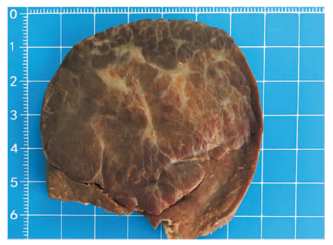
| 病例 | 性别 | 年龄(岁) | 最大径(cm) | 脾脏标本 重量(g) | 主诉 | 影像学检查CT平扫+增强 | 随访 |
|---|---|---|---|---|---|---|---|
| 1 | 女 | 35 | 7.0 | 800 | 体检发现脾脏占位1周 | 无 | 无复发 |
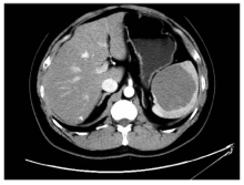
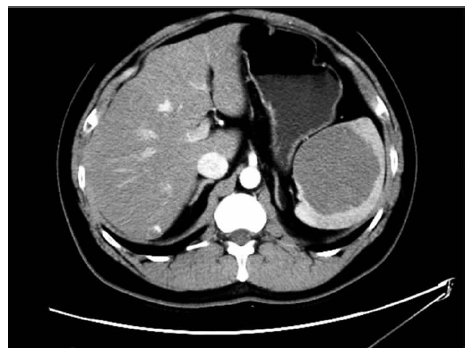
| 2 | 男 | 24 | 5.5 | 174 | 体检发现脾脏占位3 d | 脾内见类圆形稍低密度影,增强后轻度强化 | 无复发 |
| 3 | 男 | 32 | 11 | 650 | 8年前体检发现脾脏占位 | 脾脏见多个类圆形低密度影,增强后未见明显强化, 门静脉期和延迟期造影剂向肿块中心填充 | 无复发 |
| 4 | 女 | 67 | 4.2 | 590 | 20 d前因上腹痛检查 发现脾脏占位 | 脾内见类圆形稍低密度影,增强后轻度强化 | 无复发 |
| 5 | 男 | 40 | 9.0 | 410 | 体检发现脾脏占位1周 | 脾脏可见一类圆形稍低密度影,增强期后动脉期 未见明显强化,静脉期其内可见小片状强化 | 无复发 |
| 6 | 男 | 3 | 5.5 | 130 | 左上腹部扪及一包块7 d | 无 | 无复发 |
| 7 | 女 | 51 | 1.5 | 110 | 体检发现脾脏占位8 d | 脾脏近脾门处见类圆形稍低密度影,边界不清, 增强见轻度强化 | 无复发 |
| 8 | 女 | 14 | 5.5 | 210 | 反复右下腹痛1个月余,体 检发现脾脏占位1个月余 | 脾可见稍低密度影,边界欠清,呈分叶状, 增强后动脉见条索状强化 | 无复发 |

| [1] |
Falk GA, Nooli NP, Morris-Stiff G, et al. Sclerosing Angiomatoid Nodular Transformation (SANT) of the spleen: Case report and review of the literature[J]. Int J Surg Case Rep, 2012, 3(10):492-500.
doi: 10.1016/j.ijscr.2012.06.003 URL |
| [2] |
Martel M, Cheuk W, Lombardi L, et al. Sclerosing angiomatoid nodular transformation (SANT): report of 25 cases of a distinctive benign splenic lesion[J]. Am J Surg Pathol, 2004, 28(10):1268-1279.
doi: 10.1097/01.pas.0000138004.54274.d3 URL |
| [3] | Agrawal M, Uppin SG, Bh S, et al. Sclerosing Angiomatoid Nodular Transformation of the Spleen: A New Entity or a New Name?[J]. Turk Patoloji Derg, 2016, 32(3):205-210. |
| [4] | Gaeta R, Donati F, Kauffmann EF, et al. A Splenic IgG4+ Sclerosing Angiomatoid Nodular Transformation (SANT) Treated by Hemisplenectomy: A Radiologic, Histochemical, and Immunohistochemical Study[J/OL]. Appl Immunohistochem Mol Morphol, 2017-08-02[2019-06-25]. https://www.ncbi.nlm.nih.gov/pubmed/28777155. |
| [5] |
Weinreb I, Bailey D, Battaglia D, et al. CD30 and Epstein-Barr virus RNA expression in sclerosing angiomatoid nodular transformation of spleen[J]. Virchows Arch, 2007, 451(1):73-79.
pmid: 17492312 |
| [6] |
Diebold J, Le Tourneau A, Marmey B, et al. Is sclerosing angiomatoid nodular transformation (SANT) of the splenic red pulp identical to inflammatory pseudotumour? Report of 16 cases[J]. Histopathology, 2008, 53(3):299-310.
doi: 10.1111/j.1365-2559.2008.03101.x pmid: 18643852 |
| [7] | Atas H, Bulus H, Akkurt G. Sclerosing Angiomatoid Nodular Transformation of the Spleen: An uncommon Cause of Abdominal Pain[J]. Euroasian J Hepatogastroenterol, 2017, 7(1):89-91. |
| [8] |
Demirci I, Kinkel H, Antoine D, et al. Sclerosing angiomatoid nodular transformation of the spleen mimicking metastasis of melanoma: a case report and review of the literature[J]. J Med Case Rep, 2017, 11(1):251.
doi: 10.1186/s13256-017-1400-6 URL |
| [9] | 杜煜, 时高峰, 王亚宁, 等. 脾硬化性血管瘤样结节性转化的CT表现并文献复习[J]. 放射学实践, 2017, 32(2):171-174. |
| [10] |
Sharma P. 18F-FDG avid Sclerosing Angiomatoid Nodular Transformation (SANT) of spleen on PET-CT - a rare mimicker of metastasis[J]. Nucl Med Rev Cent East Eur, 2018, 21(1):53.
doi: 10.5603/NMR.2018.0014 pmid: 29442349 |
| [11] |
Wang TB, Hu BG, Liu DW, et al. Sclerosing angiomatoid nodular transformation of the spleen: A case report and literature review[J]. Oncol Lett, 2016, 12(2):928-932.
doi: 10.3892/ol.2016.4720 URL |
| [12] | Cipolla C, Florena AM, Ferrara G, et al. Sclerosing Angiomatoid Nodular Transformation: Laparoscopic Splenectomy as Therapeutic and Diagnostic Approach at the Same Time[J]. Case Rep Surg, 2018, 2018:7020538. |
| [13] | Kim KH, Lee S, Youn SH, et al. Laparoscopic splenectomy for sclerosing angiomatoid nodular transformation of the spleen[J]. J Korean Surg Soc, 2011, 80(Suppl 1):S59-S62. |
| [1] | WANG Zhaohui, WU Haibo. Clinicopathological analysis of 31 cases of gastric schwannoma [J]. Journal of Diagnostics Concepts & Practice, 2021, 20(06): 552-556. |
| [2] | CHANG Rui, XU Jiaxu, DONG Haipeng, WU Mengxiong, ZHAO Xuesong, MIAO Fei, YAN Fuhua. Value of CT spectral imaging in the evaluation of Crohn's disease activity [J]. Journal of Diagnostics Concepts & Practice, 2019, 18(04): 432-435. |
| [3] | YANG Ruxue, LI Nan, ZHOU Ting, ZHAO Yan, CHEN Shaohua, ZHU Qing, FENG Zhenzhong. Clinicopathologic analysis of skin melanocyte lesions [J]. Journal of Diagnostics Concepts & Practice, 2018, 17(05): 566-571. |
| [4] | WU Xinyang, ZHANG Huan, PAN Zilai, TAN Jingwen, GAO Xiaoyuan. The diagnostic value of dual-source CT in differentiating primary gastric lymphoma from advanced gastric cancer [J]. Journal of Diagnostics Concepts & Practice, 2018, 17(01): 60-65. |
| [5] | ZHU Peipei, ZOU Jue, CHEN Jun, XU Rongrong, YAN Hongzhu. Intracranial solitary fibrous tumor/hemangiopericytoma: a clinicopathologic study of 20 cases with review of literature [J]. Journal of Diagnostics Concepts & Practice, 2017, 16(06): 622-626. |
| [6] | YI Lin, XIAO Li, CHEN Yan, YIN Yulei. Anaplastic large cell lymphoma: a clinicopathological study and review of literature [J]. Journal of Diagnostics Concepts & Practice, 2017, 16(03): 313-319. |
| [7] | . [J]. Journal of Diagnostics Concepts & Practice, 2014, 13(05): 491-494. |
| [8] | . [J]. Journal of Diagnostics Concepts & Practice, 2013, 12(02): 170-174. |
| [9] | . [J]. Journal of Diagnostics Concepts & Practice, 2012, 11(06): 585-588. |
| [10] | . [J]. Journal of Diagnostics Concepts & Practice, 2012, 11(01): 38-41. |
| [11] | . [J]. Journal of Diagnostics Concepts & Practice, 2011, 10(06): 535-539. |
| [12] | . [J]. Journal of Diagnostics Concepts & Practice, 2011, 10(05): 428-433. |
| [13] | . [J]. Journal of Diagnostics Concepts & Practice, 2011, 10(05): 459-462. |
| [14] | . [J]. Journal of Diagnostics Concepts & Practice, 2010, 9(02): 173-176. |
| [15] | . [J]. Journal of Diagnostics Concepts & Practice, 2009, 8(03): 322-324. |
| Viewed | ||||||
|
Full text |
|
|||||
|
Abstract |
|
|||||