诊断学理论与实践 ›› 2024, Vol. 23 ›› Issue (03): 324-329.doi: 10.16150/j.1671-2870.2024.03.011
倪亚平1, 陈一峰1, 杨晓群2, 陈晓炎2,3( )
)
收稿日期:2024-03-29
接受日期:2024-04-30
出版日期:2024-06-25
发布日期:2024-06-25
通讯作者:
陈晓炎 E-mail:554531007@qq.com基金资助:
NI Yaping1, CHEN Yifeng1, YANG Xiaoqun2, CHEN Xiaoyan2,3()
Received:2024-03-29
Accepted:2024-04-30
Published:2024-06-25
Online:2024-06-25
摘要:
目的:探讨原发性肺腺癌伴肠母细胞分化(lung adenocarcinoma with enteroblastic differentiation, LAED)的临床病理特征及鉴别诊断要点。方法:回顾性分析2018年至2022年搜集的2例LAED患者的临床及影像学资料、病理形态学、免疫表型特征及基因检测结果等,并复习相关文献。结果:2例患者均为中老年男性,且长期吸烟,血清甲胎蛋白(alpha-fetoprotein,AFP)水平分别为>20 000 ng/mL、914.17 ng/mL;病灶分别位于右肺上叶及左肺下叶,最大径为12.5 cm及4.0 cm。患者的手术切除标本可见,肿瘤切面呈灰白、灰红,为实性,质软,局部易碎。镜下见大部分肿瘤组织呈实性,少部分呈腺管状、乳头状或呈囊腺样生长;肿瘤细胞学的细胞质透亮,富含糖原。免疫组化检测可见,肿瘤组织同时具有胚胎性分化和肠型分化的表型,不表达肝细胞分化标志。分子检测显示,EGFR、ALK/ROS1、RET、KRAS,BRAF、NTRK及MET均未见突变,HER-2未见扩增,EBER阴性。2例患者均诊断为LAED,因形态学及免疫表型有交叉,且均可有AFP水平升高,易被误诊为肺肝样腺癌及其他具有透明胞质的低分化腺癌。1例患者放弃治疗,于诊断2个月后去世;另1例患者接受根治性肺叶切除,术后行辅助化疗及免疫、靶向治疗,治疗后AFP水平降至正常,随访40个月时,患者因发生肿瘤骨、脑转移去世。结论:LAED目前国际上尚未见报道,其诊断及鉴别诊断主要依赖特征性的组织结构及细胞形态,并结合免疫组化标志物及血清AFP水平。本报道拓宽了产AFP的原发肺腺癌的疾病谱,LAED总体上临床进展快,患者预后差。
中图分类号:
倪亚平, 陈一峰, 杨晓群, 陈晓炎. 原发性肺腺癌伴肠母细胞分化2例临床病理及预后分析[J]. 诊断学理论与实践, 2024, 23(03): 324-329.
NI Yaping, CHEN Yifeng, YANG Xiaoqun, CHEN Xiaoyan. Primary lung adenocarcinoma with enteroblastic differentiation: a clinicopathological and prognostic analysis of two cases[J]. Journal of Diagnostics Concepts & Practice, 2024, 23(03): 324-329.

图1
肺部增强CT图像 A:例1患者,纵隔窗,左上角肺窗,见右肺上叶巨大占位;B:例2患者,肺窗,左上角纵隔窗,左肺下叶见不规则团块影。
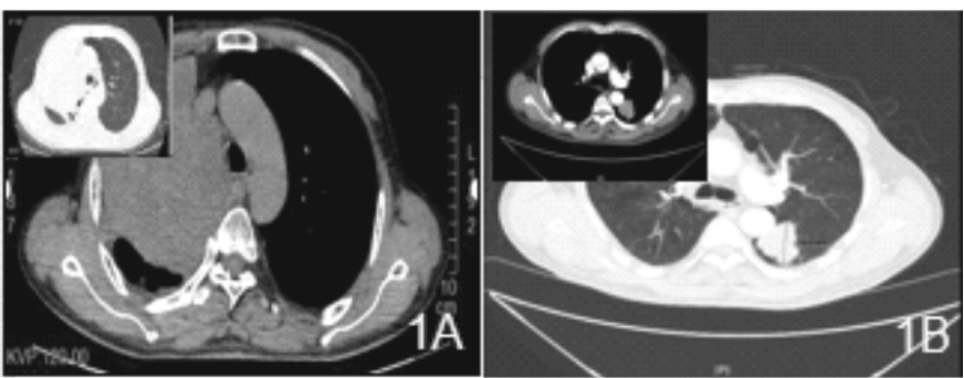

图2
例1患者的病理学图片 A:低倍镜(×40)下,肿瘤细胞大部分呈实性片状生长伴灶性坏死,少部分呈腺管、乳头状。B、C、D为中倍镜(×200)下。B:实性区肿瘤细胞立方,细胞质透明,富含糖原(AB-PAS证实);C:部分区域肿瘤细胞呈腺管、乳头状结构,细胞立方,细胞质透明,间质富含薄壁毛细血管;D:实性区域部分肿瘤细胞质内可见嗜伊红物质。E:高倍镜(×400)下,肿瘤细胞实性片状生长,细胞立方,细胞质透明。

图3
例2患者的病理学图片 A:低倍镜(×40)下,肿瘤细胞大部分实性片状生长伴灶性坏死,少部分呈腺管状、囊腺样生长;B:中倍镜(×200)下,肿瘤细胞大部分呈腺管状(左上侧),小部分呈实性片状生长(右下),细胞立方,细胞质透明,富含糖原;C:瘤细胞呈囊腺状样生长伴灶性坏死;D:实性区域细胞立方,细胞质透明,富含糖原,间质富含薄壁血管。


图4
免疫组化染色图片(EnVision两步法) Immunohistochemical staining image(EnVision two-step method) A-C:例1,肿瘤细胞表达GPC-3、AFP、SATB-2(×200);D-E:例2,2个区域肿瘤细胞表达均SALL4、Villin(×200)。

| [1] |
KITADA M, OZAWA K, SATO K, et al. Alpha-fetoprotein-producing primary lung carcinoma: a case report[J]. World J Surg Oncol, 2011, 9:47.
doi: 10.1186/1477-7819-9-47 pmid: 21554678 |
| [2] | SHORE K T, PHELPS K C, BALANI J, et al. Alpha-Fetoprotein-Producing Esophageal Adenocarcinoma With Enteroblastic, Yolk Sac Tumor-Like, and Hepatoid Carcinoma Differentiation: A Rare Case and Literature Review[J]. Int J Surg Pathol, 2023, 31(5):884-889. |
| [3] | XIAO F L, GUO Q Z, WEI H, et al. High-grade fetal adenocarcinoma of the lung with abnormal expression of alpha-fetoprotein in a female patient: Case report[J]. Medicine (Baltimore), 2021, 100(7):e24634. |
| [4] |
HIROSHIMA K, IYODA A, TOYOZAKI T, et al. Alpha-fetoprotein-producing lung carcinoma: report of three cases[J]. Pathol Int, 2002, 52(1):46-53.
doi: 10.1046/j.1440-1827.2002.01311.x pmid: 11940206 |
| [5] | WHO Classification of Tumours Editorial Board. Thoracic Tumours[M]. 5th ed. Lyon: iarc press, 2021. |
| [6] |
MURAKAMI T, YAO T, MITOMI H, et al. Clinicopathologic and immunohistochemical characteristics of gastric adenocarcinoma with enteroblastic differentiation: a study of 29 cases[J]. Gastric Cancer, 2016, 19(2):498-507.
doi: 10.1007/s10120-015-0497-9 pmid: 25893262 |
| [7] | 汪琪, 张岩, 谭聪, 等. 伴肠母细胞分化的结直肠腺癌8例临床病理学分析[J]. 中华病理学杂志, 2024, 53(4):370-376. |
| WANG Q, ZHANG Y, TAN C, et al. Colorectal adenocarcinoma with enteroblastic differentiation: a clinicopathological analysis of eight cases[J]. Chin J Pathol, 2024, 53(4):370-376. | |
| [8] | RODRÍGUEZ-VILLENA A, VELIZ-DOMÍNGUEZ A, GONZÁLEZ-GARCÍA I, et al. Enteroblastic adenocarcinoma of the ampulla of Vater[J]. Rev Esp Patol, 2024, 57(2):151-155. |
| [9] | THAKORE-SHAH K, KOLEILAT T, JAN M, et al. REST/NRSF Knockdown Alters Survival, Lineage Differentiation and Signaling in Human Embryonic Stem Cells[J]. PLoS One, 2015, 10(12):e0145280. |
| [10] | HOU Z, XIE J, ZHANG L, et al. Hepatoid Adenocarcinoma of the Lung: A Systematic Review of the Literature From 1981 to 2020[J]. Front Oncol, 2021, 11:702216. |
| [11] | WANG Y, WEI X, KE B, et al. Exploring the molecular characteristics of the malignant potential of gastric adenocarcinoma with enteroblastic differentiation[J]. Histopathology, 2023, 83(4):631-646. |
| [1] | 张祥钦, 江勇. 宏基因组第二代测序技术诊断鹦鹉热衣原体肺炎1例[J]. 诊断学理论与实践, 2022, 21(05): 635-637. |
| [2] | 谢吻, 梁怀予, 董磊, 袁菲, 王朝夫, 郭滟. 胰腺导管腺癌重要驱动基因突变与临床病理特征、预后间相关性的分析[J]. 诊断学理论与实践, 2022, 21(05): 581-587. |
| [3] | 王昭晖, 吴海波. 胃神经鞘瘤31例临床病理学分析[J]. 诊断学理论与实践, 2021, 20(06): 552-556. |
| [4] | 李娟, 刘劲松, 李梅, 李殿炜, 朱弘. 细支气管腺瘤10例临床病理分析及文献复习[J]. 诊断学理论与实践, 2021, 20(05): 466-470. |
| [5] | 吴冬梅, 吴丽莉, 陈佳, 刘坤. 淋巴上皮样肝细胞肝癌一例报告附文献复习[J]. 诊断学理论与实践, 2021, 20(05): 498-501. |
| [6] | 杨一娴, 倪仲馨, 夏蜀珺, 周伟, 詹维伟. 多灶性与单灶性甲状腺乳头状癌的临床病理特征及超声表现的比较[J]. 诊断学理论与实践, 2021, 20(02): 168-172. |
| [7] | 韦若蕖, 余红, 姚志荣. 儿童成纤维细胞结缔组织痣一例报道并文献复习[J]. 诊断学理论与实践, 2021, 20(02): 190-194. |
| [8] | 余舒文, 方正滢, 谢静远. 基因检测在慢性肾脏病诊治中的应用及进展[J]. 诊断学理论与实践, 2020, 19(06): 613-617. |
| [9] | 孟磊俊, 张晶, 王雪莉, 李治, 张泓, 曾乃燕. 儿童伯基特淋巴瘤中差异表达基因的鉴定及临床应用[J]. 诊断学理论与实践, 2020, 19(03): 248-257. |
| [10] | 何燕燕, 冯砅锦, 蔚青. 前列腺多形性巨细胞腺癌一例报告及文献复习[J]. 诊断学理论与实践, 2019, 18(2): 160-164. |
| [11] | 王志威, 张晓晓, 王杰, 魏敏, 邵玉国, 籍敏, 杨莉, 何奇. 局部晚期乳腺癌患者腋窝淋巴结转移范围的影响因素分析[J]. 诊断学理论与实践, 2019, 18(2): 189-192. |
| [12] | 金娇莺, 李倩玉, 蒋虹伟, 韩冬艳, 奚豪, 蔚青. 混合性嗜铬细胞瘤1例报道并文献复习[J]. 诊断学理论与实践, 2019, 18(2): 165-169. |
| [13] | 王顺利, 邓双双, 高慧, 肖天羽, 高金莉. 乳腺包裹性乳头状癌的临床和病理特征分析[J]. 诊断学理论与实践, 2019, 18(1): 89-92. |
| [14] | 刘立伟, 杨晓群, 范德生. 宫颈肝样腺癌一例报告及文献复习[J]. 诊断学理论与实践, 2019, 18(06): 680-682. |
| [15] | 韩冬艳, 付慧君, 何燕燕, 奚豪, 蔚青. 内淋巴囊肿瘤临床病理分析及文献复习[J]. 诊断学理论与实践, 2018, 17(06): 711-714. |
| 阅读次数 | ||||||
|
全文 |
|
|||||
|
摘要 |
|
|||||