诊断学理论与实践 ›› 2025, Vol. 24 ›› Issue (02): 125-134.doi: 10.16150/j.1671-2870.2025.02.003
王梦真, 鲍守钰, 刘鹏, 严福华, 杨文洁( )
)
收稿日期:2024-12-10
接受日期:2025-03-08
出版日期:2025-04-25
发布日期:2025-07-11
通讯作者:
杨文洁 E-mail:lisa_ywj@163.com基金资助:
WANG Mengzhen, BAO Shouyu, LIU Peng, YAN Fuhua, YANG Wenjie()
Received:2024-12-10
Accepted:2025-03-08
Published:2025-04-25
Online:2025-07-11
摘要:
光子计数CT(photon-counting computed tomography, PCCT)是近十年来CT成像领域的一项革命性技术突破,相较于传统的能量积分探测器CT,PCCT是在探测器层面对单光子水平进行成像,具备更高的空间分辨率、更少的伪影及更精准的多能谱成像,在心血管疾病诊断方面展现出极大的应用前景,尤其是线束硬化伪影的降低和超高分辨率的实现,可进一步提高冠脉狭窄评估的特异度和阳性预测值,对支架内管腔再狭窄的精准评估、斑块成分的识别及易损斑块的识别表征也得益于此。PCCT可在低辐射剂量下获得稳定的钙化积分,且虚拟非增强算法支持在增强图像中获得可靠的钙化积分,有助于进一步降低辐射剂量。PCCT在冠脉周围脂肪影像组学分析中能够提高特征可重复性,虚拟非增强算法可准确评估心外膜脂肪体积并显著降低辐射剂量;PCCT在高时间分辨率下获取的能谱图像支持单期心肌细胞外容积分数(extracellular volume, ECV)测量,也可为经导管主动脉瓣植入/置换术术前规划及术后复查提供多维度解剖信息和功能参数。尽管PCCT在冠心病诊断及心肌组织定量分析中潜力巨大,其定量结果仍受重建参数(如卷积核、虚拟单能量水平、迭代等级)影响,目前尚缺乏统一的标准和多中心研究的验证,且高分辨率模式的辐射剂量增加等问题仍限制了其广泛应用。未来应进一步开展大样本、多中心前瞻性研究,优化成像参数、标准化后处理流程,并结合人工智能工具以提升PCCT在心血管疾病诊断中的临床应用价值。
中图分类号:
王梦真, 鲍守钰, 刘鹏, 严福华, 杨文洁. 光子计数CT在心血管疾病中的应用[J]. 诊断学理论与实践, 2025, 24(02): 125-134.
WANG Mengzhen, BAO Shouyu, LIU Peng, YAN Fuhua, YANG Wenjie. Application of photon-counting CT in cardiovascular diseases[J]. Journal of Diagnostics Concepts & Practice, 2025, 24(02): 125-134.
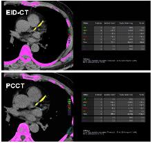
图1
PCCT和EID-CT钙化积分示意图注:65岁男性患者,EID-CT管电压120 kV,层厚3 mm,Agatston分数为958,CT 剂量指数体积(CTDIvol)为2.23 mGy;PCCT管电压120 kV,70 keV重建,层厚3 mm,Agatston分数为1 102.7,CTDIvol为1.05 mGy。
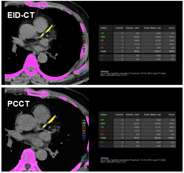
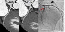
图2
PCCT能量成像去钙化图像注:虚拟单能量图像显示左回旋支近端钙化斑块狭窄率70%,去钙化图像对应30%狭窄,经ICA证实该钙化斑块造成30%管腔狭窄。
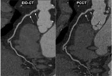
图3
PCCT和EID-CT对不同成分斑块的显示对比图注:66岁男性患者,PCCT对右冠状动脉近段的非钙化斑块及钙化斑块的边界显示较EID-CT清晰。

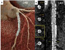
图4
63岁男性患者的冠状动脉左前降支支架PCCT图像注:PCCT可清晰地显示支架的形态结构及支架内膜增生。图像获取采用双源PCCT扫描仪,层厚0.2 mm,FOV 200 mm,kernel为Bv76,重建分辨率矩阵1 024,QIR 4级。A:三维容积再现成像;B:支架内管腔轴位测量;C:曲面重组图像。
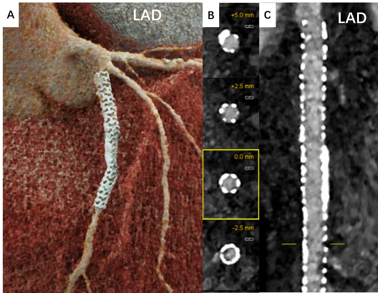
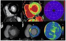
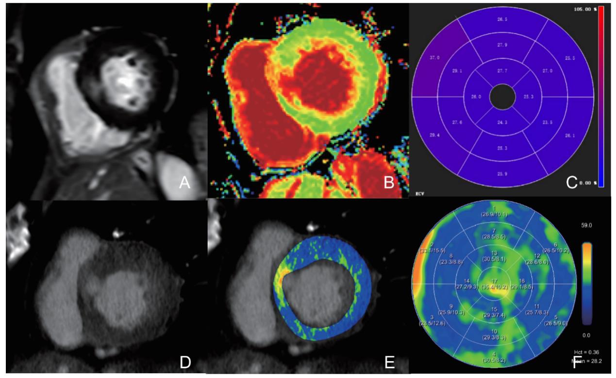
图5
PCCT与CMR延迟强化及细胞外容积定量对比注:63岁女性,重度主动脉瓣狭窄,CT-ECV(28.2%)与CMR-ECV(27.0%)的空间分布及绝对定量值均高度匹配。A:基底短轴位延迟钆增强心血管磁共振(LGE-CMR)图像,显示室间隔中部强化;B-C:对应短轴位CMR-ECV图(基于美国心脏协会17节段模型);D:基底短轴位CT延迟碘图,显示相同受累区域;E-F:对应短轴位CT-ECV图(同17节段模型)。
| [1] |
HSIEH S S, LENG S, RAJENDRAN K, et al. Photon counting CT: clinical applications and future developments[J]. IEEE Trans Radiat Plasma Med Sci,2021,5(4):441-452.
doi: 10.1109/trpms.2020.3020212 pmid: 34485784 |
| [2] |
ANDREINI D, MUSHTAQ S, PONTONE G, et al. CT perfusion versus coronary CT angiography in patients with suspected in-stent restenosis or CAD progression[J]. JACC Cardiovasc Imaging,2020,13(3):732-742.
doi: S1936-878X(19)30590-X pmid: 31422127 |
| [3] |
VANHECKE T E, MADDER R D, WEBER J E, et al. Development and validation of a predictive screening tool for uninterpretable coronary CT angiography results[J]. Circ Cardiovasc Imaging,2011,4(5):490-497.
doi: 10.1161/CIRCIMAGING.111.964205 pmid: 21775504 |
| [4] | RIJLAARSDAM-HERMSEN D, LO-KIOENG-SHIOE M S, KUIJPERS D, et al. Prognostic value of the coronary artery calcium score in suspected coronary artery disease: a study of 644 symptomatic patients[J]. Neth Heart J,2020,28(1):44-50. |
| [5] | WANG M, ZHANG X, LI J, et al. Quantification accuracy in photon-counting detector CT for coronary artery calcium score: a pilot study[J]. Int J Cardiovasc Imaging,2024,40(10):2181-2191. |
| [6] |
DOBROLINSKA M M, VAN DER WERF N R, VAN DER BIE J, et al. Radiation dose optimization for photon-counting CT coronary artery calcium scoring for different patient sizes: a dynamic phantom study[J]. Eur Radiol,2023,33(7):4668-4675.
doi: 10.1007/s00330-023-09434-1 pmid: 36729174 |
| [7] | MERGEN V, HIGASHIGAITO K, ALLMENDINGER T, et al. Tube voltage-independent coronary calcium scoring on a first-generation dual-source photon-counting CT-a proof-of-principle phantom study[J]. Int J Cardiovasc Imaging,2022,38(4):905-912. |
| [8] | EBERHARD M, MERGEN V, HIGASHIGAITO K, et al. Coronary calcium scoring with first generation dual-source photon-counting CT-first evidence from phantom and in-vivo scans[J]. Diagnostics (Basel),2021,11(9):1708. |
| [9] |
VAN DER WERF N R, GREUTER M J W, BOOIJ R, et al. Coronary calcium scores on dual-source photon-counting computed tomography: an adapted Agatston methodology aimed at radiation dose reduction[J]. Eur Radiol,2022,32(8):5201-5209.
doi: 10.1007/s00330-022-08642-5 pmid: 35230517 |
| [10] | EMRICH T, AQUINO G, SCHOEPF U J, et al. Coronary computed tomography angiography-based calcium sco-ring: In vitro and in vivo validation of a novel Virtual no-niodine reconstruction algorithm on a clinical, first-generation dual-source photon counting-detector system[J]. Invest Radiol,2022,57(8):536-543. |
| [11] | FINK N, ZSARNOCZAY E, SCHOEPF U J, et al. Photon counting detector CT-based virtual noniodine reconstruction algorithm for in vitro and in vivo coronary artery calcium scoring: impact of virtual monoenergetic and quantum iterative reconstructions[J]. Invest Radiol,2023,58(9):673-680. |
| [12] | FINK N, EMRICH T, SCHOEPF U J, et al. Improved detection of small and low-density plaques in virtual noncontrast imaging-based calcium scoring at photon-counting detector CT[J]. Radiol Cardiothorac Imaging, 2024,6(4):e230328. |
| [13] | KOTRONIAS R A, DE MARIA G L, XIE C, et al. Benchmarking photon-counting computed tomography angiography against invasive assessment of coronary stenosis: implications for severely calcified coronaries[J]. JACC Cardiovasc Imaging,2025,18(5):572-582. |
| [14] | HAGAR M T, SOSCHYNSKI M, SAFFAR R, et al. Accuracy of ultrahigh-resolution photon-counting CT for detecting coronary artery disease in a high-risk population[J]. Radiology,2023,307(5):e223305. |
| [15] | SAKAI K, SHIN D, SINGH M, et al. Diagnostic performance and clinical impact of photon-counting detector computed tomography in coronary artery disease[J]. J Am Coll Cardiol,2025,85(4):339-348. |
| [16] | HALFMANN M C, BOCKIUS S, EMRICH T, et al. Ultrahigh-spatial-resolution photon-counting detector CT angiography of coronary artery disease for stenosis assessment[J]. Radiology,2024,310(2):e231956. |
| [17] | VECSEY-NAGY M, TREMAMUNNO G, SCHOEPF U J, et al. Intraindividual Comparison of ultrahigh-spatial-resolution photon-counting detector CT and energy-integrating detector ct for coronary stenosis measurement[J]. Circ Cardiovasc Imaging,2024,17(10):e017112. |
| [18] | WOLF E V, HALFMANN M C, VARGA-SZEMES A, et al. Photon-counting detector CT virtual monoenergetic images for coronary artery stenosis quantification: Phantom and in vivo evaluation[J]. Am J Roentgenol,2024,222(3):e2330481. |
| [19] |
SARTORETTI T, MOSER L J, RUSEK S, et al. Photon-counting detector coronary CT angiography: Defining the optimal monoenergetic level for grading of calcified coronary stenosis[J]. J Cardiovasc Comput Tomogr,2024,18(6):616-617.
doi: 10.1016/j.jcct.2024.06.007 pmid: 38876897 |
| [20] | NISHIHARA T, MIYOSHI T, NAKASHIMA M, et al. Diagnostic improvements of calcium-removal image reconstruction algorithm using photon-counting detector CT for calcified coronary lesions[J]. Eur J Radiol,2024,172:111354. |
| [21] | MERGEN V, RUSEK S, CIVAIA F, et al. Virtual calcium removal in calcified coronary arteries with photon-counting detector CT-first in-vivo experience[J]. Front Cardiovasc Med,2024,11:1367463. |
| [22] |
ALLMENDINGER T, NOWAK T, FLOHR T, et al. Photon-counting detector CT-based vascular calcium removal algorithm: assessment using a cardiac motion phantom[J]. Invest Radiol,2022,57(6):399-405.
doi: 10.1097/RLI.0000000000000853 pmid: 35025834 |
| [23] | SI-MOHAMED S A, BOCCALINI S, LACOMBE H, et al. Coronary CT angiography with photon-counting CT: first-in-human results[J]. Radiology,2022,303(2):303-313. |
| [24] | ROTZINGER D C, RACINE D, BECCE F, et al. Performance of spectral photon-counting coronary CT angiography and comparison with energy-integrating-detector CT: Objective assessment with model observer[J]. Diagnostics (Basel),2021,11(12):2376. |
| [25] | MERGEN V, EBERHARD M, MANKA R, et al. First in-human quantitative plaque characterization with ultra-high resolution coronary photon-counting CT angiography[J]. Front Cardiovasc Med,2022,9:981012. |
| [26] | VECSEY-NAGY M, TREMAMUNNO G, SCHOEPF U J, et al. Coronary plaque quantification with ultrahigh-spatial-resolution photon-counting detector CT: Intraindividual comparison with energy-integrating detector CT[J]. Radiology,2025,314(3):e241479. |
| [27] | EMRICH T, HELL M. Plaque composition on ultra-high-resolution coronary computed tomography angiography with optical coherence tomography correlation[J]. Eur Heart J,2023,44(19):1765. |
| [28] | PATEL M R, NØRGAARD B L, FAIRBAIRN T A, et al. 1-Year impact on medical practice and clinical outcomes of FFRCT: The ADVANCE registry[J]. JACC Cardiovasc Imaging,2020,13(1 Pt 1):97-105. |
| [29] | AYX I, LICHTI L, BUETTNER S, et al. Feasibility of on-site CT-FFR analysis on cardiac photon-counting CT in evaluation of hemodynamically significant stenosis in comparison to invasive catheter angiography[J]. Eur J Radiol,2025,183:111927. |
| [30] | ZSARNOCZAY E, PINOS D, SCHOEPF U J, et al. Intrain-dividual comparison of coronary CT angiography-based FFR between energy-integrating and photon-counting detector CT systems[J]. Int J Cardiol,2024,399:131684. |
| [31] | VECSEY-NAGY M, TREMAMUNNO G, SCHOEPF U J, et al. Coronary CT angiography-based FFR with ultrahigh-resolution photon-counting detector CT: Intra-individual comparison to energy-integrating detector CT[J]. Eur J Radiol,2024,181:111797. |
| [32] | MELONI A, MAFFEI E, POSITANO V, et al. Technical principles, benefits, challenges, and applications of photon counting computed tomography in coronary imaging: a narrative review[J]. Cardiovasc Diagn Ther,2024,14(4):698-724. |
| [33] | 张挽时. 光子计数CT成像技术和临床价值[J]. 中华放射学杂志,2023,57(10):1133-1136. |
| ZHANG W S. Imaging technique and clinical value of photon counting CT[J]. Chin J Radiol,2023,57(10): 1133-1136. | |
| [34] |
MANNIL M, HICKETHIER T, VON SPICZAK J, et al. Photon-counting CT: High-resolution imaging of coronary stents[J]. Invest Radiol,2018,53(3):143-149.
doi: 10.1097/RLI.0000000000000420 pmid: 28945655 |
| [35] | BOCCALINI S, SI-MOHAMED S A, LACOMBE H, et al. First in-human results of computed tomography angiog-raphy for coronary stent assessment with a spectral photon counting computed tomography[J]. Invest Radiol,2022,57(4):212-221. |
| [36] | FAHRNI G, BOCCALINI S, PRIEUR C, et al. Comprehensive imaging of coronary stent using ultra-high resolution spectral photon counting CT: A multimodality validation[J]. JACC Cardiovasc Interv,2023,16(19):2466-2468. |
| [37] | PETRITSCH B, PETRI N, WENG A M, et al. Photon-counting computed tomography for coronary stent ima-ging: in vitro evaluation of 28 coronary stents[J]. Invest Radiol,2021,56(10):653-660. |
| [38] |
SYMONS R, DE BRUECKER Y, ROOSEN J, et al. Quarter-millimeter spectral coronary stent imaging with photon-counting CT: Initial experience[J]. J Cardiovasc Comput Tomogr,2018,12(6):509-515.
doi: S1934-5925(18)30426-X pmid: 30509378 |
| [39] | RAJAGOPAL J R, FARHADI F, RICHARDS T, et al. Evaluation of coronary plaques and stents with conventional and photon-counting CT: Benefits of high-resolution photon-counting CT[J]. Radiol Cardiothorac Imaging,2021,3(5):e210102. |
| [40] | DECKER J A, O'DOHERTY J, SCHOEPF U J, et al. Stent imaging on a clinical dual-source photon-counting detector CT system-impact of luminal attenuation and sharp kernels on lumen visibility[J]. Eur Radiol,2023,33(4):2469-2477. |
| [41] | KOONS E K, THORNE J E, HUBER N R, et al. Quanti-fying lumen diameter in coronary artery stents with high-resolution photon counting detector CT and convolutional neural network denoising[J]. Med Phys,2023,50(7):4173-4181. |
| [42] | QIN L, ZHOU S, DONG H, et al. Improvement of coronary stent visualization using ultra-high-resolution photon-counting detector CT[J]. Eur Radiol,2024,34(10):6568-6577. |
| [43] | STEIN T, VON ZUR MUHLEN C, VERLOH N, et al. Evaluating small coronary stents with dual-source photon-counting computed tomography: effect of different scan modes on image quality and performance in a phantom[J]. Diagn Interv Radiol,2025,31(1):29-38. |
| [44] |
BRATKE G, HICKETHIER T, BAR-NESS D, et al. Spectral photon-counting computed tomography for coronary stent imaging: Evaluation of the potential clinical impact for the delineation of in-stent restenosis[J]. Invest Radiol,2020,55(2):61-67.
doi: 10.1097/RLI.0000000000000610 pmid: 31524765 |
| [45] | ELIAS MICHAEL A, SCHOENBECK D, MICHAEL WOELTJEN M, et al. Photon counting computed tomography of in-stent-stenosis in a phantom: Optimal virtual monoenergetic imaging in ultra high resolution[J]. Heliyon,2024,10(6):e27636. |
| [46] | HAGAR M T, SOSCHYNSKI M, SAFFAR R, et al. Ultra-high-resolution photon-counting detector CT in evaluati-ng coronary stent patency: a comparison to invasive coronary angiography[J]. Eur Radiol,2024,34(7):4273-4283. |
| [47] | KRAVCHENKO D, VECSEY-NAGY M, TREMAMUNNO G, et al. Intra-individual comparison of epicardial adipose tissue characteristics on coronary CT angiography between photon-counting detector and energy-integrating detector CT systems[J]. Eur J Radiol,2024,181:111728. |
| [48] | MERGEN V, RIED E, ALLMENDINGER T, et al. Epicardial adipose tissue attenuation and fat attenuation index: phantom study and in vivo measurements with photon-counting detector CT[J]. AJR Am J Roentgenol,2022,218(5):822-829. |
| [49] | KRAVCHENKO D, VECSEY-NAGY M, VARGA-SZEMES A, et al. Intra-individual radiomic analysis of pericoronary adipose tissue: Photon-counting detector vs energy-integrating detector CT angiography[J]. Int J Cardiol,2025,420:132749. |
| [50] |
TREMAMUNNO G, VECSEY-NAGY M, HAGAR M T, et al. Intra-individual differences in pericoronary fat attenuation index measurements between photon-counting and energy-integrating detector computed tomography[J]. Acad Radiol,2025,32(3):1333-1343.
doi: 10.1016/j.acra.2024.11.055 pmid: 39665893 |
| [51] | RISCH F, SCHWARZ F, BRAUN F, et al. Assessment of epicardial adipose tissue on virtual non-contrast images derived from photon-counting detector coronary CTA datasets[J]. Eur Radiol,2023,33(4):2450-2460. |
| [52] | KAHMANN J, NÖRENBERG D, PAPAVASSILIU T, et al. Combined conventional factors and the radiomics signature of coronary plaque texture could improve cardiac risk prediction[J]. Insights Imaging,2024,15(1):170. |
| [53] | LISI C, KLAMBAUER K, MOSER L J, et al. The pericoronary adipose tissue attenuation in CT strongly depends on kernels and iterative reconstructions[J]. Eur Radiol,2025,35(5):2866-2876. |
| [54] |
NACIF M S, KAWEL N, LEE J J, et al. Interstitial myocardial fibrosis assessed as extracellular volume fraction with low-radiation-dose cardiac CT[J]. Radiology,2012,264(3):876-883.
doi: 10.1148/radiol.12112458 pmid: 22771879 |
| [55] |
BANDULA S, WHITE S K, FLETT A S, et al. Measurement of myocardial extracellular volume fraction by using equilibrium contrast-enhanced CT: validation against histologic findings[J]. Radiology,2013,269(2):396-403.
doi: 10.1148/radiol.13130130 pmid: 23878282 |
| [56] |
JABLONOWSKI R, WILSON M W, DO L, et al. Multidetector CT measurement of myocardial extracellular volume in acute patchy and contiguous infarction: validation with microscopic measurement[J]. Radiology,2015,274(2):370-378.
doi: 10.1148/radiol.14140131 pmid: 25247406 |
| [57] | LEE H J, IM D J, YOUN J C, et al. Myocardial extracellular volume fraction with dual-energy equilibrium contrast-enhanced cardiac CT in nonischemic cardio-myopathy: A prospective comparison with cardiac MR imaging[J]. Radiology,2016,280(1):49-57. |
| [58] |
MERGEN V, SARTORETTI T, KLOTZ E, et al. Extracellular volume quantification with cardiac late enhancement scanning using dual-source photon-counting detector CT[J]. Invest Radiol,2022,57(6):406-411.
doi: 10.1097/RLI.0000000000000851 pmid: 35066531 |
| [59] | AQUINO G J, O'DOHERTY J, SCHOEPF U J, et al. Myocardial characterization with extracellular volume mapping with a first-generation photon-counting detector CT with MRI reference[J]. Radiology,2023,307(2):e222030. |
| [60] |
ODA S, FUNAMA Y, KOJIMA S, et al. Basic verification of myocardial extracellular volume quantification by prototype photon-counting detector computed tomography: A phantom study[J]. J Clin Imaging Sci,2025,15:8.
doi: 10.25259/JCIS_157_2024 pmid: 40041436 |
| [61] |
GNASSO C, PINOS D, SCHOEPF U J, et al. Impact of reconstruction parameters on the accuracy of myocardial extracellular volume quantification on a first-generation, photon-counting detector CT[J]. Eur Radiol Exp,2024,8(1):70.
doi: 10.1186/s41747-024-00469-7 pmid: 38890175 |
| [62] | GKIZAS C, LONGERE B, SLIWICKA O, et al. Photon-counting CT-derived extracellular volume in acute myocarditis: Comparison with cardiac MRI[J]. Diagn Interv Imaging,2025. |
| [63] | MERGEN V, EHRBAR N, MOSER L J, et al. Synthetic hematocrit from virtual non-contrast images for myocardial extracellular volume evaluation with photon-counting detector CT[J]. Eur Radiol,2024,34(12):7845-7855. |
| [64] | DIRRICHS T, SCHRÖDER J, FRICK M, et al. Photon-counting versus dual-source CT for transcatheter aortic valve implantation planning[J]. Acad Radiol,2024,31(12):4780-4789. |
| [65] | BRENDEL J M, WALTERSPIEL J, HAGEN F, et al. Coronary artery disease evaluation during transcatheter aortic valve replacement work-up using photon-counting CT and artificial intelligence[J]. Diagn Interv Imaging,2024,105(7-8):273-280. |
| [66] | RIPPEL K, DECKER J A, LUITJENS J, et al. Virtual monoenergetic imaging of thoracoabdominal computed tomography angiography on photon-counting detector computertomography: Assessment of image quality and leveraging low-keV series for salvaging suboptimal contrast acquisitions[J]. Diagnostics (Basel),2024,14(24):2843. |
| [67] | RIPPEL K, LUITJENS J, HABEEBALLAH O, et al. Evaluation of ECG-gated, high-pitch thoracoabdominal angiographies with dual-source photon-counting detector computed tomography[J]. J Endovasc Ther,2024. |
| [68] |
RISCH F, HARMEL E, RIPPEL K, et al. Virtual non-contrast series of photon-counting detector computed tomography angiography for aortic valve calcium scoring[J]. Int J Cardiovasc Imaging,2024,40(4):723-732.
doi: 10.1007/s10554-023-03040-4 pmid: 38175389 |
| [1] | 黄瑞坤, 杨琰昭, 柴维敏. 光子计数CT在胰腺成像中的应用进展[J]. 诊断学理论与实践, 2025, 24(02): 111-117. |
| [2] | 李卫侠, 严福华. 光子计数CT在肝脏疾病中的应用进展[J]. 诊断学理论与实践, 2025, 24(02): 118-124. |
| [3] | 蔡欣欣, 邓嵘, 徐欣欣, 许芷涵, 常蕊, 董海鹏, 林慧敏, 严福华. 基于光子计数CT的肝脏脂肪分数定量测定与磁共振质子密度脂肪分数间的一致性研究[J]. 诊断学理论与实践, 2025, 24(02): 146-154. |
| [4] | 常蕊, 李纪强, 杨琰昭, 柴维敏, 严福华, 董海鹏. 光子计数CT胰腺低剂量动态容积灌注扫描中单期图像对胰腺癌图像的评估价值[J]. 诊断学理论与实践, 2025, 24(02): 155-162. |
| [5] | 周山税, 秦乐, 常蕊, 杜联军, 严福华, 刘方韬. 基于光子计数探测器CT能谱定位像定量评估股骨颈骨密度的前瞻性研究[J]. 诊断学理论与实践, 2025, 24(02): 163-169. |
| [6] | 吕海英, 陆勇, 贺娜英. 光子计数CT在神经系统成像中的临床价值[J]. 诊断学理论与实践, 2025, 24(02): 212-219. |
| [7] | 范婧, 杨文洁, 王梦真, 陆伟, 石骁萌, 朱宏. 深度学习重建算法在低管电压冠状动脉CT血管成像中的应用[J]. 诊断学理论与实践, 2022, 21(03): 374-379. |
| [8] | 施仲伟. 回眸过去30年全球和中国的心血管疾病负担及其危险因素——1990年至2019年全球心血管疾病负担及其危险因素报告解读[J]. 诊断学理论与实践, 2021, 20(04): 349-355. |
| [9] | 吴歆, 耿旭强, 徐沪济. 多基因风险评分在复杂性状疾病中的应用进展[J]. 诊断学理论与实践, 2020, 19(05): 540-543. |
| [10] | 倪瀚文, 吴立群. 外泌体在心肌缺血及房颤诊治中的应用前景研究进展[J]. 诊断学理论与实践, 2020, 19(02): 199-202. |
| [11] | 陈瑶瑶, 顾爱华. 氧化三甲胺与心血管疾病关系的研究进展[J]. 诊断学理论与实践, 2019, 18(2): 237-240. |
| [12] | 华沙, 赵建荣,. 载脂蛋白A5调节三酰甘油代谢及其对心血管疾病影响[J]. 诊断学理论与实践, 2014, 13(03): 336-340. |
| [13] | 庞小芬, 高丽红,. 老年骨质疏松症与心血管疾病相关性的认识[J]. 诊断学理论与实践, 2012, 11(01): 15-18. |
| [14] | 方怡, 陈芳源,. 组织因子微粒的研究进展[J]. 诊断学理论与实践, 2011, 10(06): 563-566. |
| [15] | 张一帆, 尹红燕,. 干细胞治疗缺血性心脏病分子显像的机遇和挑战[J]. 诊断学理论与实践, 2011, 10(01): 18-21. |
| 阅读次数 | ||||||
|
全文 |
|
|||||
|
摘要 |
|
|||||