诊断学理论与实践 ›› 2023, Vol. 22 ›› Issue (02): 190-196.doi: 10.16150/j.1671-2870.2023.02.014
陈乾, 林慧敏, 严福华( )
)
收稿日期:2023-01-11
出版日期:2023-04-25
发布日期:2023-08-31
通讯作者:
严福华 E-mail:yfh11655@rjh.com.cn
基金资助:
CHEN Qian, LIN Huimin, YAN Fuhua()
Received:2023-01-11
Online:2023-04-25
Published:2023-08-31
摘要:
肝功能储备在肝脏疾病在术前评估中意义重大。早先,由于技术限制,很少有研究探索磁共振成像(magnatic resonance imaging, MRI)在肝脏功能储备定量中的应用;近年来MRI不断发展和完善,在成像方式、造影材料、定量参数等多个方向获得进展,产生了多种用于定量肝功能的新技术、新应用。MRI弹性成像技术可以反映生物力学特征,通过振动波在肝内传播速率等参数,可以得到肝脏微结构与功能的信息;肝胆细胞特异性对比剂增强MRI可以被肝细胞特异性摄取,从而显示肝细胞的功能情况,并在MRI图像上直观地显示出肝脏的功能分布;T1弛豫时间成像以及扩散加权成像等技术则能够反映大分子成分与水扩散等局部微环境特点,并通过这些特征定量肝功能。此外,诸如三维断层弹性成像以及结合T1 mapping定量的肝胆细胞特异性对比剂增强MRI等最新技术进展进一步提高了这些检查定量评估肝功能的效能。与临床常用的生化指标、Child-Pugh评分以及吲哚菁绿试验等肝功能定量方法相比,影像学检查虽仍然受制于成本高、操作困难等因素,但其提供的的肝功能储备空间分布信息可协助肝脏术前规划,从而更好地预测术后不良事件,提高患者的生存概率。所以,影像学技术在肝功能定量中有着很好的发展前景。
中图分类号:
陈乾, 林慧敏, 严福华. 磁共振成像评估肝功能储备的研究进展[J]. 诊断学理论与实践, 2023, 22(02): 190-196.
CHEN Qian, LIN Huimin, YAN Fuhua. Advances in the evaluation of hepatic function by magnetic resonance imaging[J]. Journal of Diagnostics Concepts & Practice, 2023, 22(02): 190-196.
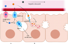
图1
对比剂的代谢路径示意图 传统的磁共振对比剂(黑色球)不能被肝细胞摄取。A:钆塞酸二钠的代谢示意图。钆塞酸二钠(蓝色球)与吲哚青绿或胆红素等物质共用部分代谢通路,通过受体OATP1进入肝细胞,并最终排泄到胆管中。B:99mTc-GSA的代谢示意图。99mTc-GSA(黄色球)结合肝细胞膜上ASPGR受体,然后在肝细胞中被内吞、消化和分解。由于结合同一受体的物质之间通常具有竞争性抑制作用,这种代谢途径的差异可能对检查方法的应用场景有很大的影响。

| [1] | 国家卫生健康委办公厅. 原发性肝癌诊疗指南(2022年版)[J]. 中华外科杂志, 2022, 60(4):273-309. |
| General Office of the National Health Commission. Guidelines for diagnosis and treatment of primary liver cancer(The 2022 edition)[J]. Chin J Surg, 2022, 60(4):273-309. | |
| [2] |
Zheng R, Zhang S, Zeng H, et al. Cancer incidence and mortality in China, 2016[J]. J Natl Cancer Center, 2022, 2(1):1-9.
doi: 10.1016/j.jncc.2022.02.002 URL |
| [3] | RAHNEMAI-AZAR A A, CLOYD J M, WEBER S M, et al. Update on Liver Failure Following Hepatic Resection: Strategies for Prediction and Avoidance of Post-operative Liver Insufficiency[J]. J Clin Transl Hepatol, 2018, 6(1): 97-104. |
| [4] |
VENKATESH S K, YIN M, EHMAN R L. Magnetic resonance elastography of liver: technique, analysis, and clinical applications[J]. J Magn Reson Imaging, 2013, 37(3):544-555.
doi: 10.1002/jmri.23731 pmid: 23423795 |
| [5] |
SINGH S, VENKATESH S K, WANG Z, et al. Diagnostic performance of magnetic resonance elastography in sta-ging liver fibrosis: a systematic review and meta-analysis of individual participant data[J]. Clin Gastroenterol Hepatol, 2015, 13(3):440-451,e6.
doi: 10.1016/j.cgh.2014.09.046 URL |
| [6] |
KARIN D, KOYAMA Y, BRENNER D, et al. The characteristics of activated portal fibroblasts/myofibroblasts in liver fibrosis[J]. Differentiation, 2016, 92(3):84-92.
doi: S0301-4681(15)30100-6 pmid: 27591095 |
| [7] |
VENKATESH S K, WELLS M L, MILLER F H, et al. Magnetic resonance elastography: beyond liver fibrosis-a case-based pictorial review[J]. Abdom Radiol (NY), 2018, 43(7):1590-1611.
doi: 10.1007/s00261-017-1383-1 pmid: 29143076 |
| [8] |
KUSAKA K, HARIHARA Y, TORZILLI G, et al. Objective evaluation of liver consistency to estimate hepatic fibrosis and functional reserve for hepatectomy[J]. J Am Coll Surg, 2000, 191(1):47-53.
doi: 10.1016/S1072-7515(00)00309-4 URL |
| [9] |
LI B, MIN J, LIANG W R, et al. Use of magnetic resonance elastography for assessing liver functional reserve: A clinical study[J]. World J Gastroenterol, 2015, 21(24):7522-7528.
doi: 10.3748/wjg.v21.i24.7522 URL |
| [10] |
LIN H, WANG Y, ZHOU J, et al. Tomoelastography based on multifrequency MR elastography predicts liver function reserve in patients with hepatocellular carcinoma: a prospective study[J]. Insights Imaging, 2022, 13(1):95.
doi: 10.1186/s13244-022-01232-5 |
| [11] |
HOFFMAN D H, AYOOLA A, NICKEL D, et al. MR elastography, T1 and T2 relaxometry of liver: role in noninvasive assessment of liver function and portal hypertension[J]. Abdom Radiol (NY), 2020, 45(9):2680-2687.
doi: 10.1007/s00261-020-02432-7 pmid: 32274552 |
| [12] |
HOODESHENAS S, YIN M, VENKATESH S K. Magnetic Resonance Elastography of Liver: Current Update[J]. Top Magn Reson Imaging, 2018, 27(5):319-333.
doi: 10.1097/RMR.0000000000000177 pmid: 30289828 |
| [13] |
LIU L, YOU Z, YU H, et al. Mechanotransduction-modulated fibrotic microniches reveal the contribution of angiogenesis in liver fibrosis[J]. Nat Mater, 2017, 16(12):1252-1261.
doi: 10.1038/nmat5024 pmid: 29170554 |
| [14] |
ZHANG Y N, FOWLER K J, OZTURK A, et al. Liver fibrosis imaging: A clinical review of ultrasound and magnetic resonance elastography[J]. J Magn Reson Imaging, 2020, 51(1):25-42.
doi: 10.1002/jmri.26716 pmid: 30859677 |
| [15] |
WANG J, WANG Q, YU G, et al. Correlation Between Liver Stiffness Measured by Shear Wave Elastography and Child-Pugh Classification[J]. J Ultrasound Med, 2018, 37(9):2191-2199.
doi: 10.1002/jum.14569 pmid: 29476558 |
| [16] |
HEUCKE N, WUENSCH T, MOHR J, et al. Non-invasive structure-function assessment of the liver by 2D time-harmonic elastography and the dynamic Liver MAximum capacity (LiMAx) test[J]. J Gastroenterol Hepatol, 2019, 34(9):1611-1619.
doi: 10.1111/jgh.v34.9 URL |
| [17] |
IMAJO K, HONDA Y, KOBAYASHI T, et al. Direct Comparison of US and MR Elastography for Staging Liver Fibrosis in Patients With Nonalcoholic Fatty Liver Disease[J]. Clin Gastroenterol Hepatol, 2022, 20(4):908-917,e11.
doi: 10.1016/j.cgh.2020.12.016 URL |
| [18] |
European Association for the Study of the Liver. EASL Clinical Practice Guidelines on non-invasive tests for evaluation of liver disease severity and prognosis - 2021 update[J]. J Hepatol, 2021, 75(3):659-689.
doi: 10.1016/j.jhep.2021.05.025 URL |
| [19] |
VAN BEERS B E, PASTOR C M, HUSSAIN H K. Primovist, Eovist: what to expect?[J]. J Hepatol, 2012, 57(2):421-429.
doi: 10.1016/j.jhep.2012.01.031 pmid: 22504332 |
| [20] |
FREITAS P S, JANICAS C, VEIGA J, et al. Imaging evaluation of the liver in oncology patients: A comparison of techniques[J]. World J Hepatol, 2021, 13(12):1936-1955.
doi: 10.4254/wjh.v13.i12.1936 pmid: 35069999 |
| [21] |
DAHLSTRÖM N, PERSSON A, ALBIIN N, et al. Contrast-enhanced magnetic resonance cholangiography with Gd-BOPTA and Gd-EOB-DTPA in healthy subjects[J]. Acta Radiol, 2007, 48(4):362-368.
doi: 10.1080/02841850701196922 pmid: 17453513 |
| [22] |
YOON J H, LEE J M, PAEK M, et al. Quantitative assessment of hepatic function: modified look-locker inversion recovery (MOLLI) sequence for T1 mapping on Gd-EOB-DTPA-enhanced liver MR imaging[J]. Eur Radiol, 2016, 26(6):1775-1782.
doi: 10.1007/s00330-015-3994-7 pmid: 26373756 |
| [23] |
YOON J H, LEE J M, KANG H J, et al. Quantitative Assessment of Liver Function by Using Gadoxetic Acid-enhanced MRI: Hepatocyte Uptake Ratio[J]. Radiology, 2019, 290(1):125-133.
doi: 10.1148/radiol.2018180753 pmid: 30375932 |
| [24] |
SANDRASEGARAN K, CUI E, ELKADY R, et al. Can functional parameters from hepatobiliary phase of gadoxetate MRI predict clinical outcomes in patients with cirrhosis?[J]. Eur Radiol, 2018, 28(10):4215-4224.
doi: 10.1007/s00330-018-5366-6 pmid: 29651764 |
| [25] |
WIBMER A, PRUSA A M, NOLZ R, et al. Liver failure after major liver resection: risk assessment by using preoperative Gadoxetic acid-enhanced 3-T MR imaging[J]. Radiology, 2013, 269(3):777-786.
doi: 10.1148/radiol.13130210 pmid: 23942606 |
| [26] |
LUO N, HUANG X, JI Y, et al. A functional liver ima-ging score for preoperative prediction of liver failure after hepatocellular carcinoma resection[J]. Eur Radiol, 2022, 32(8):5623-5632.
doi: 10.1007/s00330-022-08656-z |
| [27] |
SALERNO M, JANARDHANAN R, JIJI R S, et al. Comparison of methods for determining the partition coefficient of gadolinium in the myocardium using T1 mapping[J]. J Magn Reson Imaging, 2013, 38(1):217-224.
doi: 10.1002/jmri.23875 pmid: 23197434 |
| [28] |
DAHLQVIST LEINHARD O, DAHLSTRÖM N, KIHLBERG J, et al. Quantifying differences in hepatic uptake of the liver specific contrast agents Gd-EOB-DTPA and Gd-BOPTA: a pilot study[J]. Eur Radiol, 2012, 22(3):642-653.
doi: 10.1007/s00330-011-2302-4 pmid: 21984449 |
| [29] |
HAIMERL M, VERLOH N, ZEMAN F, et al. Gd-EOB-DTPA-enhanced MRI for evaluation of liver function: Comparison between signal-intensity-based indices and T1 relaxometry[J]. Sci Rep, 2017, 7:43347.
doi: 10.1038/srep43347 pmid: 28266528 |
| [30] |
BESA C, BANE O, JAJAMOVICH G, et al. 3D T1 relaxo-metry pre and post gadoxetic acid injection for the assessment of liver cirrhosis and liver function[J]. Magn Reson Imaging, 2015, 33(9):1075-1082.
doi: 10.1016/j.mri.2015.06.013 URL |
| [31] |
GEISEL D, LÜDEMANN L, HAMM B, et al. Imaging-Based Liver Function Tests--Past, Present and Future[J]. Rofo, 2015, 187(10):863-871.
doi: 10.1055/s-0035-1553306 pmid: 26230140 |
| [32] | 王荣福, 庞小溪, 刘敏, 等. 99mTc-GSA肝受体显像在肝功能评估临床研究应用及进展[J]. 世界华人消化杂志, 2017, 25(21):1903-1909. |
|
WANG R F, PANG X X, LIU M, et al. Clinical application of 99mTc-GSA in assessment of liver function by hepatic receptor imaging[J]. World Chin J Digestol, 2017, 25(21):1903-1909.
doi: 10.11569/wcjd.v25.i21.1903 URL |
|
| [33] |
KUDO M, TODO A, IKEKUBO K, et al. Functional hepatic imaging with receptor-binding radiopharmaceutical: clinical potential as a measure of functioning hepatocyte mass[J]. Gastroenterol Jpn, 1991, 26(6):734-741.
pmid: 1662653 |
| [34] | BENNINK R J, DINANT S, ERDOGAN D, et al. Preope-rative assessment of postoperative remnant liver function using hepatobiliary scintigraphy[J]. J Nucl Med, 2004, 45(6):965-971. |
| [35] |
ERDOGAN D, HEIJNEN B H, BENNINK R J, et al. Preoperative assessment of liver function: a comparison of 99mTc-Mebrofenin scintigraphy with indocyanine green clearance test[J]. Liver Int, 2004, 24(2):117-123.
doi: 10.1111/j.1478-3231.2004.00901.x pmid: 15078475 |
| [36] |
NAKAGAWA M, NAMIMOTO T, SHIMIZU K, et al. Measuring hepatic functional reserve using T1 mapping of Gd-EOB-DTPA enhanced 3T MR imaging: A preliminary study comparing with 99mTc GSA scintigraphy and signal intensity based parameters[J]. Eur J Radiol, 2017, 92:116-123.
doi: 10.1016/j.ejrad.2017.05.011 URL |
| [37] | RONG P, FENG Z, GUO R, et al. CT-based estimation of liver function using arterial enhancement fraction in liver cirrhosis patients[J]. Zhong Nan Da Xue Xue Bao Yi Xue Ban, 2019, 44(5):469-476. |
| [38] |
DE GRAAF W, BENNINK R J, HEGER M, et al. Quantitative assessment of hepatic function during liver regeneration in a standardized rat model[J]. J Nucl Med, 2011, 52(2):294-302.
doi: 10.2967/jnumed.110.078360 URL |
| [39] | NILSSON H, BLOMQVIST L, DOUGLAS L, et al. Gd-EOB-DTPA-enhanced MRI for the assessment of liver function and volume in liver cirrhosis[J]. Br J Radiol, 2013, 86(1026):20120653. |
| [40] |
HUANG M, SHEN S, CAI H, et al. Regional liver function analysis with gadoxetic acid-enhanced MRI and virtual hepatectomy: prediction of postoperative short-term outcomes for HCC[J]. Eur Radiol, 2021, 31(7):4720-4730.
doi: 10.1007/s00330-020-07606-x pmid: 33449173 |
| [41] | CLEMÉNT O, MÜHLER A, VEXLER V S, et al. Comparison of Gd-EOB-DTPA and Gd-DTPA for contrast-enhanced MR imaging of liver tumors[J]. J Magn Reson Ima-ging, 1993, 3(1):71-77. |
| [42] |
IMAI Y, KATAYAMA K, HORI M, et al. Prospective Comparison of Gd-EOB-DTPA-Enhanced MRI with Dynamic CT for Detecting Recurrence of HCC after Radiofrequency Ablation[J]. Liver Cancer, 2017, 6(4):349-359.
doi: 10.1159/000481416 pmid: 29234638 |
| [43] |
UNAL E, IDILMAN I S, KARÇAALTINCABA M. Multiparametric or practical quantitative liver MRI: towards millisecond, fat fraction, kilopascal and function era[J]. Expert Rev Gastroenterol Hepatol, 2017, 11(2):167-182.
doi: 10.1080/17474124.2017.1271710 URL |
| [44] |
DING Y, RAO S X, CHEN C, et al. Assessing liver function in patients with HBV-related HCC: a comparison of T1 mapping on Gd-EOB-DTPA-enhanced MR imaging with DWI[J]. Eur Radiol, 2015, 25(5):1392-1398.
doi: 10.1007/s00330-014-3542-x URL |
| [45] |
ZHANG J, GUO Y, TAN X, et al. MRI-based estimation of liver function by intravoxel incoherent motion diffusion-weighted imaging[J]. Magn Reson Imaging, 2016, 34(8):1220-1225.
doi: 10.1016/j.mri.2016.05.013 pmid: 27262828 |
| [46] |
CHEN F, CHEN Y L, CHEN T W, et al. Liver lobe based intravoxel incoherent motion diffusion weighted imaging in hepatitis B related cirrhosis: Association with child-pugh class and esophageal and gastric fundic varices[J]. Medicine (Baltimore), 2020, 99(2):e18671.
doi: 10.1097/MD.0000000000018671 URL |
| [47] |
WANG L, REGATTE R R. T₁ρ MRI of human musculoskeletal system[J]. J Magn Reson Imaging, 2015, 41(3): 586-600.
doi: 10.1002/jmri.24677 pmid: 24935818 |
| [48] |
RAUSCHER I, EIBER M, GANTER C, et al. Evaluation of T1ρ as a potential MR biomarker for liver cirrhosis: comparison of healthy control subjects and patients with liver cirrhosis[J]. Eur J Radiol, 2014, 83(6):900-904.
doi: S0720-048X(14)00111-9 pmid: 24661616 |
| [49] |
TAKAYAMA Y, NISHIE A, ASAYAMA Y, et al. T1 ρ Relaxation of the liver: A potential biomarker of liver function[J]. J Magn Reson Imaging, 2015, 42(1):188-195.
doi: 10.1002/jmri.24739 pmid: 25155810 |
| [50] |
SINGH A, REDDY D, HARIS M, et al. T1ρ MRI of healthy and fibrotic human livers at 1.5 T[J]. J Transl Med, 2015, 13:292.
doi: 10.1186/s12967-015-0648-0 URL |
| [1] | 李卫侠, 徐学勤, 朱晓雷, 陈克敏. 39例肾上腺皮质癌患者的CT、MRI影像特点及其诊断价值[J]. 诊断学理论与实践, 2023, 22(02): 134-140. |
| [2] | 黄娟, 朱晓雷, 李晓, 陈克敏, 严福华, 徐学勤. 血氧水平依赖磁共振成像评估早期慢性肾病肾缺氧的研究[J]. 诊断学理论与实践, 2022, 21(03): 385-389. |
| [3] | 朱乃懿, 姜奕歆, 柴丽, 柴维敏. 磁共振对超声阴性而乳腺X线检出BI-RADS4类以上钙化灶的诊断价值分析[J]. 诊断学理论与实践, 2021, 20(05): 439-444. |
| [4] | 何碧媛, 周毓青, 姚秉彝, 曹力, 包丽. 中孕期弹性超声成像宫颈机能智能定量分析预测自发性早产的临床应用价值[J]. 诊断学理论与实践, 2021, 20(05): 450-455. |
| [5] | 张雪坤, 李彦, 严福华, 赵洪飞, 宋琦. 基于光梭成像的新型加速技术在颅脑MRI中的应用价值研究[J]. 诊断学理论与实践, 2021, 20(04): 378-383. |
| [6] | 孙甜甜, 叶宝英, 杨钰, 牛建梅. 彩色多普勒超声与磁共振成像在凶险型前置胎盘及合并胎盘植入产前诊断中的应用及漏诊分析[J]. 诊断学理论与实践, 2021, 20(02): 173-177. |
| [7] | 何碧媛, 周毓青. 三维超声、超声造影及超声弹性成像在妇科疾病诊断中的应用进展及策略[J]. 诊断学理论与实践, 2020, 19(06): 626-629. |
| [8] | 周伟, 侯怡卿, 詹维伟. 超声造影及超声弹性成像在良恶性甲状腺结节鉴别诊断中的应用进展[J]. 诊断学理论与实践, 2020, 19(04): 344-349. |
| [9] | 吴霜, 解骞, 管雪妮, 张素芳, 高信芳, 梁宗辉. 磁共振体素内不相干运动扩散加权成像诊断活动期克罗恩病的价值及效能分析[J]. 诊断学理论与实践, 2020, 19(02): 157-161. |
| [10] | 唐桢云, 詹维伟. 剪切波弹性成像在乳腺癌诊断中的应用现状[J]. 诊断学理论与实践, 2019, 18(2): 223-227. |
| [11] | 曹烨, 刘晓晟, 葛晓乾, 周斌. 运用动态增强磁共振成像评估颈动脉粥样斑块稳定性的初步研究[J]. 诊断学理论与实践, 2019, 18(04): 436-441. |
| [12] | 季沁, 周一帆, 陈茉, 李杰, 丁文波, 钱涛, 褚晓秋, 王建华, 徐书杭, 刘超. 弹性成像联合ACR-TIRADS诊断甲状腺结节良恶性的临床价值研究[J]. 诊断学理论与实践, 2019, 18(03): 307-312. |
| [13] | 朱晓雷, 陈璐, 陆文丽, 刘燕, 严福华, 王伟, 董治亚. 474例中枢性性早熟女童不同年龄段垂体MRI影像学异常比例分析[J]. 诊断学理论与实践, 2019, 18(03): 286-290. |
| [14] | 李云峰, 江泓, 李宁, 孙青芳. 核磁共振成像诊断三叉神经痛的价值分析与研究[J]. 诊断学理论与实践, 2018, 17(05): 562-565. |
| [15] | 许晶晶, 张敏鸣. 人工智能机器学习方法在阿尔茨海默病中的应用现状[J]. 诊断学理论与实践, 2018, 17(04): 466-470. |
| 阅读次数 | ||||||
|
全文 |
|
|||||
|
摘要 |
|
|||||