诊断学理论与实践 ›› 2023, Vol. 22 ›› Issue (02): 134-140.doi: 10.16150/j.1671-2870.2023.02.005
李卫侠, 徐学勤( ), 朱晓雷, 陈克敏
收稿日期:2023-05-22
出版日期:2023-04-25
发布日期:2023-08-31
通讯作者:
徐学勤 E-mail:xxq11550@rjh.com.cn
LI Weixia, XU Xueqin(), ZHU Xiaolei, CHEN Kemin
Received:2023-05-22
Online:2023-04-25
Published:2023-08-31
摘要:
目的:分析肾上腺皮质腺癌(adrenocortical carcinoma, ACC)的CT、MRI表现特点,并探讨二者诊断ACC的价值。方法:回顾分析经手术及病理检查证实的39例ACC患者的CT、MRI平扫及增强检查表现及临床资料,总结对ACC有较大诊断价值的影像学征象,比较2种成像技术在ACC诊断中的价值。结果:14例患者同时完成了CT和MRI平扫加增强检查,22例患者仅接受了CT平扫加增强检查,3例患者仅接受了MRI检查。39例ACC患者中,38例肿瘤为单侧发病(左侧19例、右侧19例),1例为双侧发病;23例肿瘤呈分叶状,16例呈类圆形;肿瘤直径为4.0~17.3 cm。CT、MRI检查中,分别有47.2%、82.4%的病例病灶内存在出血灶;91.7%、50%的病例发现肿瘤内增粗、迂曲的肿瘤血管影;77.8%、82.4%的病例提示包膜不光整。所有肿瘤均呈不均匀强化,肿瘤实性成分多呈中度增强;少量病例可见钙化、周围脏器血管受累、腹膜后淋巴结及远处肿瘤转移。14例同时完成CT和MRI平扫加增强检查的病例中,2种成像技术分别发现肿瘤灶内出血6例、12例,肿瘤内增生血管12例、6例,包膜不光整9例、12例。结论:ACC的CT、MRI表现具有一定特征性,多表现为体积较大(直径多大于4 cm),病灶呈分叶状,内多见肿瘤血管、出血坏死,肿瘤累及包膜;增强后多呈中度不均匀强化。CT在检出肿瘤血管影方面具有一定优势,而MRI在显示肿瘤内出血方面具有一定优势。
中图分类号:
李卫侠, 徐学勤, 朱晓雷, 陈克敏. 39例肾上腺皮质癌患者的CT、MRI影像特点及其诊断价值[J]. 诊断学理论与实践, 2023, 22(02): 134-140.
LI Weixia, XU Xueqin, ZHU Xiaolei, CHEN Kemin. Characteristics and diagnostic value of CT and MRI in adrenocortical carcinoma: a study of 39 cases[J]. Journal of Diagnostics Concepts & Practice, 2023, 22(02): 134-140.
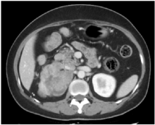
图1
增强CT示右侧肾上腺分叶状团块呈不均匀中度强化

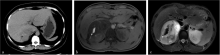
图2
ACC病灶内出血表现 a:CT平扫,见右侧肾上腺肿块内见小斑片状稍高密度灶;b、c:MRI平扫图像,右侧肾上腺肿块内见小斑片状T1WI高信号(b)、T2WI低信号(c)。

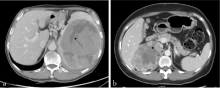
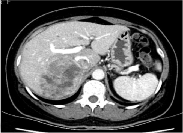
图3
CT增强图像示ACC病灶内坏死 a:肾上腺肿块内裂隙状坏死;b:囊状坏死伴壁结节。
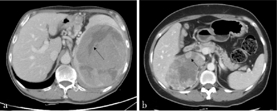

图4
CT增强图像见ACC病灶内部增粗迂曲的肿瘤血管影
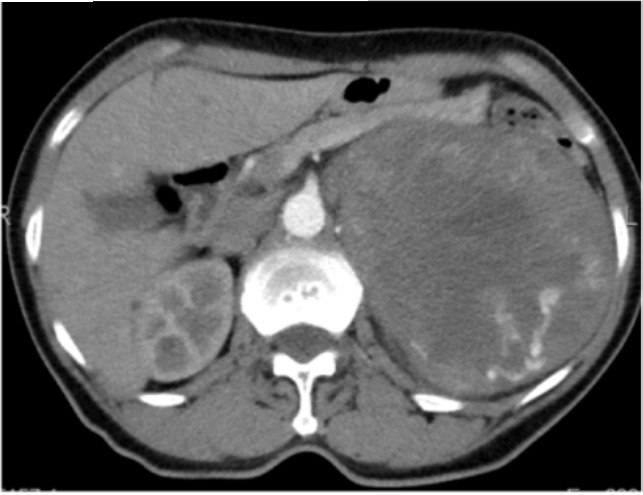
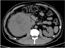
图5
CT平扫图像见右侧肾上腺肿块包膜不光整


图6
CT增强图像见右侧肾上腺不均匀强化肿块 局部强化的肿瘤栓子突入下腔静脉
| [14] |
AHMED A A, THOMAS A J, GANESHAN D M, et al. Adrenal cortical carcinoma: pathology, genomics, prognosis, imaging features, and mimics with impact on management[J]. Abdom Radiol (NY), 2020, 45(4):945-963.
doi: 10.1007/s00261-019-02371-y pmid: 31894378 |
| [15] | BENASSAI G, DESIATO V, BENASSAI G, et al. Adrenocortical carcinoma: what the surgeon needs to know. Case report and literature review[J]. Int J Surg, 2014, 12(Suppl 1):S22-S28. |
| [16] |
ZINI L, PORPIGLIA F, FASSNACHT M. Contemporary management of adrenocortical carcinoma[J]. Eur Urol, 2011, 60(5):1055-1065.
doi: 10.1016/j.eururo.2011.07.062 pmid: 21831516 |
| [17] |
ADKINS K M, LEE J T, BRESS A L, et al. Classic Cus-hing's syndrome in a patient with adrenocortical carcinoma[J]. Radiol Case Rep, 2015, 8(3):826.
doi: 10.2484/rcr.v8i3.826 URL |
| [18] |
ELSE T, KIM AC, SABOLCH A, et al. Adrenocortical carcinoma[J]. Endocr Rev, 2014, 35(2):282-326.
doi: 10.1210/er.2013-1029 pmid: 24423978 |
| [19] |
BHARWANI N, ROCKALL A G, SAHDEV A, et al. Adrenocortical carcinoma: the range of appearances on CT and MRI[J]. Am J Roentgenol, 2011, 196(6):W706-W714.
doi: 10.2214/AJR.10.5540 URL |
| [20] |
SHIN Y R, KIM K A. Imaging Features of Various Adrenal Neoplastic Lesions on Radiologic and Nuclear Medicine Imaging[J]. Am J Roentgenol, 2015, 205(3):554-563.
doi: 10.2214/AJR.15.14467 pmid: 26295641 |
| [21] |
RIBEIRO J, RIBEIRO R C, FLETCHER B D. Imaging findings in pediatric adrenocortical carcinoma[J]. Pediatr Radiol, 2000, 30(1):45-51.
pmid: 10663510 |
| [22] |
ROWE S P, LUGO-FAGUNDO C, AHN H, et al. What the radiologist needs to know: the role of preoperative computed tomography in selection of operative approach for adrenalectomy and review of operative techniques[J]. Abdom Radiol (NY), 2019, 44(1):140-153.
doi: 10.1007/s00261-018-1669-y pmid: 29967985 |
| [23] |
KAWASHIMA A, SANDLER C M, ERNST R D, et al. Imaging of nontraumatic hemorrhage of the adrenal gland[J]. Radiographics, 1999, 19(4):949-963.
doi: 10.1148/radiographics.19.4.g99jl13949 pmid: 10464802 |
| [1] |
ABIVEN G, COSTE J, GROUSSIN L, et al. Clinical and biological features in the prognosis of adrenocortical cancer: poor outcome of cortisol-secreting tumors in a series of 202 consecutive patients[J]. J Clin Endocrinol Metab, 2006, 91(7):2650-2655.
doi: 10.1210/jc.2005-2730 URL |
| [2] |
SHARMA E, DAHAL S, SHARMA P, et al. The Characteristics and Trends in Adrenocortical Carcinoma: A United States Population Based Study[J]. J Clin Med Res, 2018, 10(8):636-640.
doi: 10.14740/jocmr3503w pmid: 29977421 |
| [3] |
GRUBBS E, LEE J E. Limited prognostic value of the 2004 International Union Against Cancer staging classification for adrenocortical carcinoma: proposal for a revised TNM classification[J]. Cancer, 2009, 115(24):5847.
doi: 10.1002/cncr.24693 pmid: 19827149 |
| [4] | 金晓龙, 袁菲, 蔚青, 等. 肾上腺肿瘤和瘤样病变1166例病理分析[J]. 诊断学理论与实践, 2003, 2(2):119-121,125. |
| JIN X L, YUAN F, WEI Q, et al. Pathologically Analyzing 1166 Cases of Adrenal Tumors and Tumor-like Lesions[J]. J Diagn Concepts & Pract, 2003, 2(2):119-121,125. | |
| [5] | MANTERO F, TERZOLO M, ARNALDI G, et al. A survey on adrenal incidentaloma in Italy. Study Group on Adrenal Tumors of the Italian Society of Endocrinology[J]. J Clin Endocrinol Metab, 2000, 85(2):637-644. |
| [6] | 赵勤余, 韩志江, 陈克敏. 肾上腺皮质癌的CT诊断及鉴别诊断[J]. 放射学实践, 2012, 27(9):975-978. |
| ZHAO Q Y, HAN Z J, CHEN K M. Adrenocortical carcinoma:value of CT diagnosis and differential diagnosis[J]. Radiol Pract, 2012, 27(9):975-978. | |
| [7] | 汪建华, 丁前江, 马小龙, 等. 肾上腺原发性皮质腺癌的CT与MRI表现及其病理基础[J]. 中华放射学杂志, 2016, 50(11):882-885. |
| WANG J H, DING Q J, MA X L, et al. Primary adrenocortical carcinoma: CT and MRI evaluation with pathological correlation[J]. Chin J Radiol, 2016, 50(11):882-885. | |
| [8] | 沃方明, 王玉涛, 张建, 等. 肾上腺皮质癌的CT、MRI及PET/CT表现[J]. 医学影像学杂志, 2018, 28(6):993-996,1000. |
| WO F M, WANG Y T, ZHANG J, et al. CT, MRI and PET/CT features of adrenocortical carcinoma[J]. J Med Imaging, 2018, 28(6):993-996,1000. | |
| [9] | 茹立, 陈挺, 李盛, 等. 肾上腺皮质腺癌的CT、MR影像学特点及临床表现并文献复习[J]. 医学影像学杂志, 2019, 29(11):1985-1988. |
| RU L, CHEN T, LI S, et al. CT and MR imaging features and clinical manifestations of adrenocortical carcinoma and literature review[J]. J Med Imaging, 2019, 29(11):1985-1988. | |
| [10] | 许晓琴, 姚振威, 林含舜, 等. 原发性肾上腺皮质腺癌的CT表现与病理特点[J]. 中国医学计算机成像杂志, 2019, 25(1):37-41. |
| XU X Q, YAO Z W, LIN H S, et al. CT Manifestations and Pathological Features of Primary Adrenocortical Carcinoma[J]. Chin Comput Med Imaging, 2019, 25(1):37-41. | |
| [11] | 苏停停, 尚进, 袁佳, 等. 肾上腺皮质癌影像学表现[J]. 中国医学影像技术, 2020, 36(12):1839-1842. |
| SU T T, SHANH J, YUAN J, et al. Imaging manifestations of adrenocortical carcinoma[J]. Chin J Med Imaging Technol, 2020, 36(12):1839-1842. | |
| [12] |
PALOKA R, GOPIREDDY D R, VIRARKAR M, et al. Multimodality imaging of adrenal gland pathologies: A comprehensive pictorial review[J]. J Clin Imaging Sci, 2022, 12:62.
doi: 10.25259/JCIS_92_2022 pmid: 36601600 |
| [13] |
SHARMA E, DAHAL S, SHARMA P, et al. The Characteristics and Trends in Adrenocortical Carcinoma: A United States Population Based Study[J]. J Clin Med Res, 2018, 10(8):636-640.
doi: 10.14740/jocmr3503w pmid: 29977421 |
| [1] | 陈乾, 林慧敏, 严福华. 磁共振成像评估肝功能储备的研究进展[J]. 诊断学理论与实践, 2023, 22(02): 190-196. |
| [2] | 黄娟, 朱晓雷, 李晓, 陈克敏, 严福华, 徐学勤. 血氧水平依赖磁共振成像评估早期慢性肾病肾缺氧的研究[J]. 诊断学理论与实践, 2022, 21(03): 385-389. |
| [3] | 中华医学会内分泌学分会. 新型冠状病毒肺炎疫情下肾上腺疾病管理专家建议[J]. 诊断学理论与实践, 2022, 21(02): 139-142. |
| [4] | 朱乃懿, 姜奕歆, 柴丽, 柴维敏. 磁共振对超声阴性而乳腺X线检出BI-RADS4类以上钙化灶的诊断价值分析[J]. 诊断学理论与实践, 2021, 20(05): 439-444. |
| [5] | 张雪坤, 李彦, 严福华, 赵洪飞, 宋琦. 基于光梭成像的新型加速技术在颅脑MRI中的应用价值研究[J]. 诊断学理论与实践, 2021, 20(04): 378-383. |
| [6] | 孙甜甜, 叶宝英, 杨钰, 牛建梅. 彩色多普勒超声与磁共振成像在凶险型前置胎盘及合并胎盘植入产前诊断中的应用及漏诊分析[J]. 诊断学理论与实践, 2021, 20(02): 173-177. |
| [7] | 曹琪琪, 秦乐, 周慧娟, 杨之涛, 苏文婷, 杨文洁, 程增辉, 陆勇, 严福华, 潘自来. 新型冠状病毒(2019-nCoV)肺炎的CT征象分析[J]. 诊断学理论与实践, 2020, 19(1): 16-19. |
| [8] | 吴霜, 解骞, 管雪妮, 张素芳, 高信芳, 梁宗辉. 磁共振体素内不相干运动扩散加权成像诊断活动期克罗恩病的价值及效能分析[J]. 诊断学理论与实践, 2020, 19(02): 157-161. |
| [9] | 王兰, 张欢, 葛颖倩, 陆静, 崔征, 颜凌, 潘自来. 胃癌肝转移病灶的人工智能辅助半自动分割软件的临床应用评估[J]. 诊断学理论与实践, 2019, 18(05): 515-520. |
| [10] | 曹烨, 刘晓晟, 葛晓乾, 周斌. 运用动态增强磁共振成像评估颈动脉粥样斑块稳定性的初步研究[J]. 诊断学理论与实践, 2019, 18(04): 436-441. |
| [11] | 朱晓雷, 陈璐, 陆文丽, 刘燕, 严福华, 王伟, 董治亚. 474例中枢性性早熟女童不同年龄段垂体MRI影像学异常比例分析[J]. 诊断学理论与实践, 2019, 18(03): 286-290. |
| [12] | 韩宝惠, 沈胤晨. 我国肺癌筛查现状与展望[J]. 诊断学理论与实践, 2018, 17(05): 487-489. |
| [13] | 李云峰, 江泓, 李宁, 孙青芳. 核磁共振成像诊断三叉神经痛的价值分析与研究[J]. 诊断学理论与实践, 2018, 17(05): 562-565. |
| [14] | 许晶晶, 张敏鸣. 人工智能机器学习方法在阿尔茨海默病中的应用现状[J]. 诊断学理论与实践, 2018, 17(04): 466-470. |
| [15] | 赵华丽, 徐文鹏, 梁宗辉. 创伤性臂丛神经损伤的磁共振成像3D-FIESTA-C、IDEAL序列特征及诊断价值[J]. 诊断学理论与实践, 2018, 17(02): 197-201. |
| 阅读次数 | ||||||
|
全文 |
|
|||||
|
摘要 |
|
|||||