| [1] |
刘宝将, 朱旭, 刘鹏. 肝动脉灌注化疗在胃癌肝转移中的临床应用[J]. 中国介入影像与治疗学, 2018, 15(8):509-512.
|
| [2] |
Chen W, Zheng R, Baade PD, et al. Cancer statistics in China, 2015[J]. CA Cancer J Clin, 2016, 66(2):115-132.
doi: 10.3322/caac.21338
URL
|
| [3] |
Coccolini F, Montori G, Ceresoli M, et al. Advanced gastric cancer: What we know and what we still have to learn[J]. World J Gastroenterol, 2016, 22(3):1139-1159.
doi: 10.3748/wjg.v22.i3.1139
URL
|
| [4] |
姚强, 金俊, 邓建良, 等. 胃癌肝转移患者预后影响因素分析[J]. 肿瘤学杂志, 2018, 24(2):104-108.
|
| [5] |
潘旻, 秦日昇, 陈春桥, 等. 胃癌肝转移经导管动脉化学栓塞序贯阿帕替尼治疗的回顾性分析[J]. 临床医药文献电子杂志, 2018, 5(47):63,158.
|
| [6] |
姜彬彬, 张仲一, 严昆, 等. 经皮超声引导下射频消融治疗胃癌肝转移疗效分析[J]. 中国介入影像与治疗学, 2018, 15(1):24-28.
|
| [7] |
李相成, 邵子诚, 张嘉伟. 晚期胃癌肝转移规范化治疗策略[J]. 中国实用外科杂志, 2017, 37(10):1106-1109.
doi: 10.19538/j.cjps.issn1005-2208.2017.10.10
|
| [8] |
邹程程. 478例胃癌肝转移的临床分析[D]. 河北: 河北医科大学, 2017.
|
| [9] |
王方. 胃癌肝转移的预后因素分析[D]. 河南: 郑州大学, 2017.
|
| [10] |
陈凛, 李佶阳. 胃癌肝转移转化治疗的临床研究进展[J]. 外科理论与实践, 2017, 22(1):5-8.
|
| [11] |
徐宏智. 胃癌肝转移的外科治疗研究新进展[J]. 中国普通外科杂志, 2016, 25(10):1500-1505.
|
| [12] |
朱婷. 胃癌肝转移手术治疗的远期疗效及预后因素Meta分析[D]. 山西: 山西医科大学, 2016.
|
| [13] |
杜金轲, 黎东明. 不同方案治疗胃癌同时性肝转移的疗效比较及预后危险因素分析[J]. 中国现代普通外科进展, 2016, 19(9):691-694.
|
| [14] |
武新洋, 张欢, 潘自来, 等. 双源CT对原发性胃淋巴瘤和进展期胃癌的鉴别诊断价值[J]. 诊断学理论与实践, 2018, 17(1):61-65.
|
| [15] |
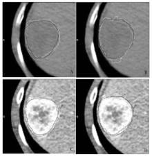
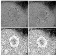
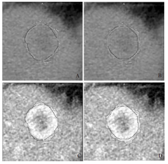
Kocak B, Durmaz ES, Kaya OK, et al. Reliability of Single-Slice-Based 2D CT Texture Analysis of Renal Masses: Influence of Intra- and Interobserver Manual Segmentation Variability on Radiomic Feature Reproducibility[J]. Am J Roentgenol, 2019, 213(2):377-383.
doi: 10.2214/AJR.19.21212
URL
|
 ), PAN Zilai4(