Journal of Diagnostics Concepts & Practice ›› 2023, Vol. 22 ›› Issue (02): 190-196.doi: 10.16150/j.1671-2870.2023.02.014
• Reviews • Previous Articles Next Articles
CHEN Qian, LIN Huimin, YAN Fuhua( )
)
Received:2023-01-11
Online:2023-04-25
Published:2023-08-31
CLC Number:
CHEN Qian, LIN Huimin, YAN Fuhua. Advances in the evaluation of hepatic function by magnetic resonance imaging[J]. Journal of Diagnostics Concepts & Practice, 2023, 22(02): 190-196.
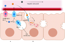
Figure 1
metabolic pathway of contrast agents

| [1] | 国家卫生健康委办公厅. 原发性肝癌诊疗指南(2022年版)[J]. 中华外科杂志, 2022, 60(4):273-309. |
| General Office of the National Health Commission. Guidelines for diagnosis and treatment of primary liver cancer(The 2022 edition)[J]. Chin J Surg, 2022, 60(4):273-309. | |
| [2] |
Zheng R, Zhang S, Zeng H, et al. Cancer incidence and mortality in China, 2016[J]. J Natl Cancer Center, 2022, 2(1):1-9.
doi: 10.1016/j.jncc.2022.02.002 URL |
| [3] | RAHNEMAI-AZAR A A, CLOYD J M, WEBER S M, et al. Update on Liver Failure Following Hepatic Resection: Strategies for Prediction and Avoidance of Post-operative Liver Insufficiency[J]. J Clin Transl Hepatol, 2018, 6(1): 97-104. |
| [4] |
VENKATESH S K, YIN M, EHMAN R L. Magnetic resonance elastography of liver: technique, analysis, and clinical applications[J]. J Magn Reson Imaging, 2013, 37(3):544-555.
doi: 10.1002/jmri.23731 pmid: 23423795 |
| [5] |
SINGH S, VENKATESH S K, WANG Z, et al. Diagnostic performance of magnetic resonance elastography in sta-ging liver fibrosis: a systematic review and meta-analysis of individual participant data[J]. Clin Gastroenterol Hepatol, 2015, 13(3):440-451,e6.
doi: 10.1016/j.cgh.2014.09.046 URL |
| [6] |
KARIN D, KOYAMA Y, BRENNER D, et al. The characteristics of activated portal fibroblasts/myofibroblasts in liver fibrosis[J]. Differentiation, 2016, 92(3):84-92.
doi: S0301-4681(15)30100-6 pmid: 27591095 |
| [7] |
VENKATESH S K, WELLS M L, MILLER F H, et al. Magnetic resonance elastography: beyond liver fibrosis-a case-based pictorial review[J]. Abdom Radiol (NY), 2018, 43(7):1590-1611.
doi: 10.1007/s00261-017-1383-1 pmid: 29143076 |
| [8] |
KUSAKA K, HARIHARA Y, TORZILLI G, et al. Objective evaluation of liver consistency to estimate hepatic fibrosis and functional reserve for hepatectomy[J]. J Am Coll Surg, 2000, 191(1):47-53.
doi: 10.1016/S1072-7515(00)00309-4 URL |
| [9] |
LI B, MIN J, LIANG W R, et al. Use of magnetic resonance elastography for assessing liver functional reserve: A clinical study[J]. World J Gastroenterol, 2015, 21(24):7522-7528.
doi: 10.3748/wjg.v21.i24.7522 URL |
| [10] |
LIN H, WANG Y, ZHOU J, et al. Tomoelastography based on multifrequency MR elastography predicts liver function reserve in patients with hepatocellular carcinoma: a prospective study[J]. Insights Imaging, 2022, 13(1):95.
doi: 10.1186/s13244-022-01232-5 |
| [11] |
HOFFMAN D H, AYOOLA A, NICKEL D, et al. MR elastography, T1 and T2 relaxometry of liver: role in noninvasive assessment of liver function and portal hypertension[J]. Abdom Radiol (NY), 2020, 45(9):2680-2687.
doi: 10.1007/s00261-020-02432-7 pmid: 32274552 |
| [12] |
HOODESHENAS S, YIN M, VENKATESH S K. Magnetic Resonance Elastography of Liver: Current Update[J]. Top Magn Reson Imaging, 2018, 27(5):319-333.
doi: 10.1097/RMR.0000000000000177 pmid: 30289828 |
| [13] |
LIU L, YOU Z, YU H, et al. Mechanotransduction-modulated fibrotic microniches reveal the contribution of angiogenesis in liver fibrosis[J]. Nat Mater, 2017, 16(12):1252-1261.
doi: 10.1038/nmat5024 pmid: 29170554 |
| [14] |
ZHANG Y N, FOWLER K J, OZTURK A, et al. Liver fibrosis imaging: A clinical review of ultrasound and magnetic resonance elastography[J]. J Magn Reson Imaging, 2020, 51(1):25-42.
doi: 10.1002/jmri.26716 pmid: 30859677 |
| [15] |
WANG J, WANG Q, YU G, et al. Correlation Between Liver Stiffness Measured by Shear Wave Elastography and Child-Pugh Classification[J]. J Ultrasound Med, 2018, 37(9):2191-2199.
doi: 10.1002/jum.14569 pmid: 29476558 |
| [16] |
HEUCKE N, WUENSCH T, MOHR J, et al. Non-invasive structure-function assessment of the liver by 2D time-harmonic elastography and the dynamic Liver MAximum capacity (LiMAx) test[J]. J Gastroenterol Hepatol, 2019, 34(9):1611-1619.
doi: 10.1111/jgh.v34.9 URL |
| [17] |
IMAJO K, HONDA Y, KOBAYASHI T, et al. Direct Comparison of US and MR Elastography for Staging Liver Fibrosis in Patients With Nonalcoholic Fatty Liver Disease[J]. Clin Gastroenterol Hepatol, 2022, 20(4):908-917,e11.
doi: 10.1016/j.cgh.2020.12.016 URL |
| [18] |
European Association for the Study of the Liver. EASL Clinical Practice Guidelines on non-invasive tests for evaluation of liver disease severity and prognosis - 2021 update[J]. J Hepatol, 2021, 75(3):659-689.
doi: 10.1016/j.jhep.2021.05.025 URL |
| [19] |
VAN BEERS B E, PASTOR C M, HUSSAIN H K. Primovist, Eovist: what to expect?[J]. J Hepatol, 2012, 57(2):421-429.
doi: 10.1016/j.jhep.2012.01.031 pmid: 22504332 |
| [20] |
FREITAS P S, JANICAS C, VEIGA J, et al. Imaging evaluation of the liver in oncology patients: A comparison of techniques[J]. World J Hepatol, 2021, 13(12):1936-1955.
doi: 10.4254/wjh.v13.i12.1936 pmid: 35069999 |
| [21] |
DAHLSTRÖM N, PERSSON A, ALBIIN N, et al. Contrast-enhanced magnetic resonance cholangiography with Gd-BOPTA and Gd-EOB-DTPA in healthy subjects[J]. Acta Radiol, 2007, 48(4):362-368.
doi: 10.1080/02841850701196922 pmid: 17453513 |
| [22] |
YOON J H, LEE J M, PAEK M, et al. Quantitative assessment of hepatic function: modified look-locker inversion recovery (MOLLI) sequence for T1 mapping on Gd-EOB-DTPA-enhanced liver MR imaging[J]. Eur Radiol, 2016, 26(6):1775-1782.
doi: 10.1007/s00330-015-3994-7 pmid: 26373756 |
| [23] |
YOON J H, LEE J M, KANG H J, et al. Quantitative Assessment of Liver Function by Using Gadoxetic Acid-enhanced MRI: Hepatocyte Uptake Ratio[J]. Radiology, 2019, 290(1):125-133.
doi: 10.1148/radiol.2018180753 pmid: 30375932 |
| [24] |
SANDRASEGARAN K, CUI E, ELKADY R, et al. Can functional parameters from hepatobiliary phase of gadoxetate MRI predict clinical outcomes in patients with cirrhosis?[J]. Eur Radiol, 2018, 28(10):4215-4224.
doi: 10.1007/s00330-018-5366-6 pmid: 29651764 |
| [25] |
WIBMER A, PRUSA A M, NOLZ R, et al. Liver failure after major liver resection: risk assessment by using preoperative Gadoxetic acid-enhanced 3-T MR imaging[J]. Radiology, 2013, 269(3):777-786.
doi: 10.1148/radiol.13130210 pmid: 23942606 |
| [26] |
LUO N, HUANG X, JI Y, et al. A functional liver ima-ging score for preoperative prediction of liver failure after hepatocellular carcinoma resection[J]. Eur Radiol, 2022, 32(8):5623-5632.
doi: 10.1007/s00330-022-08656-z |
| [27] |
SALERNO M, JANARDHANAN R, JIJI R S, et al. Comparison of methods for determining the partition coefficient of gadolinium in the myocardium using T1 mapping[J]. J Magn Reson Imaging, 2013, 38(1):217-224.
doi: 10.1002/jmri.23875 pmid: 23197434 |
| [28] |
DAHLQVIST LEINHARD O, DAHLSTRÖM N, KIHLBERG J, et al. Quantifying differences in hepatic uptake of the liver specific contrast agents Gd-EOB-DTPA and Gd-BOPTA: a pilot study[J]. Eur Radiol, 2012, 22(3):642-653.
doi: 10.1007/s00330-011-2302-4 pmid: 21984449 |
| [29] |
HAIMERL M, VERLOH N, ZEMAN F, et al. Gd-EOB-DTPA-enhanced MRI for evaluation of liver function: Comparison between signal-intensity-based indices and T1 relaxometry[J]. Sci Rep, 2017, 7:43347.
doi: 10.1038/srep43347 pmid: 28266528 |
| [30] |
BESA C, BANE O, JAJAMOVICH G, et al. 3D T1 relaxo-metry pre and post gadoxetic acid injection for the assessment of liver cirrhosis and liver function[J]. Magn Reson Imaging, 2015, 33(9):1075-1082.
doi: 10.1016/j.mri.2015.06.013 URL |
| [31] |
GEISEL D, LÜDEMANN L, HAMM B, et al. Imaging-Based Liver Function Tests--Past, Present and Future[J]. Rofo, 2015, 187(10):863-871.
doi: 10.1055/s-0035-1553306 pmid: 26230140 |
| [32] | 王荣福, 庞小溪, 刘敏, 等. 99mTc-GSA肝受体显像在肝功能评估临床研究应用及进展[J]. 世界华人消化杂志, 2017, 25(21):1903-1909. |
|
WANG R F, PANG X X, LIU M, et al. Clinical application of 99mTc-GSA in assessment of liver function by hepatic receptor imaging[J]. World Chin J Digestol, 2017, 25(21):1903-1909.
doi: 10.11569/wcjd.v25.i21.1903 URL |
|
| [33] |
KUDO M, TODO A, IKEKUBO K, et al. Functional hepatic imaging with receptor-binding radiopharmaceutical: clinical potential as a measure of functioning hepatocyte mass[J]. Gastroenterol Jpn, 1991, 26(6):734-741.
pmid: 1662653 |
| [34] | BENNINK R J, DINANT S, ERDOGAN D, et al. Preope-rative assessment of postoperative remnant liver function using hepatobiliary scintigraphy[J]. J Nucl Med, 2004, 45(6):965-971. |
| [35] |
ERDOGAN D, HEIJNEN B H, BENNINK R J, et al. Preoperative assessment of liver function: a comparison of 99mTc-Mebrofenin scintigraphy with indocyanine green clearance test[J]. Liver Int, 2004, 24(2):117-123.
doi: 10.1111/j.1478-3231.2004.00901.x pmid: 15078475 |
| [36] |
NAKAGAWA M, NAMIMOTO T, SHIMIZU K, et al. Measuring hepatic functional reserve using T1 mapping of Gd-EOB-DTPA enhanced 3T MR imaging: A preliminary study comparing with 99mTc GSA scintigraphy and signal intensity based parameters[J]. Eur J Radiol, 2017, 92:116-123.
doi: 10.1016/j.ejrad.2017.05.011 URL |
| [37] | RONG P, FENG Z, GUO R, et al. CT-based estimation of liver function using arterial enhancement fraction in liver cirrhosis patients[J]. Zhong Nan Da Xue Xue Bao Yi Xue Ban, 2019, 44(5):469-476. |
| [38] |
DE GRAAF W, BENNINK R J, HEGER M, et al. Quantitative assessment of hepatic function during liver regeneration in a standardized rat model[J]. J Nucl Med, 2011, 52(2):294-302.
doi: 10.2967/jnumed.110.078360 URL |
| [39] | NILSSON H, BLOMQVIST L, DOUGLAS L, et al. Gd-EOB-DTPA-enhanced MRI for the assessment of liver function and volume in liver cirrhosis[J]. Br J Radiol, 2013, 86(1026):20120653. |
| [40] |
HUANG M, SHEN S, CAI H, et al. Regional liver function analysis with gadoxetic acid-enhanced MRI and virtual hepatectomy: prediction of postoperative short-term outcomes for HCC[J]. Eur Radiol, 2021, 31(7):4720-4730.
doi: 10.1007/s00330-020-07606-x pmid: 33449173 |
| [41] | CLEMÉNT O, MÜHLER A, VEXLER V S, et al. Comparison of Gd-EOB-DTPA and Gd-DTPA for contrast-enhanced MR imaging of liver tumors[J]. J Magn Reson Ima-ging, 1993, 3(1):71-77. |
| [42] |
IMAI Y, KATAYAMA K, HORI M, et al. Prospective Comparison of Gd-EOB-DTPA-Enhanced MRI with Dynamic CT for Detecting Recurrence of HCC after Radiofrequency Ablation[J]. Liver Cancer, 2017, 6(4):349-359.
doi: 10.1159/000481416 pmid: 29234638 |
| [43] |
UNAL E, IDILMAN I S, KARÇAALTINCABA M. Multiparametric or practical quantitative liver MRI: towards millisecond, fat fraction, kilopascal and function era[J]. Expert Rev Gastroenterol Hepatol, 2017, 11(2):167-182.
doi: 10.1080/17474124.2017.1271710 URL |
| [44] |
DING Y, RAO S X, CHEN C, et al. Assessing liver function in patients with HBV-related HCC: a comparison of T1 mapping on Gd-EOB-DTPA-enhanced MR imaging with DWI[J]. Eur Radiol, 2015, 25(5):1392-1398.
doi: 10.1007/s00330-014-3542-x URL |
| [45] |
ZHANG J, GUO Y, TAN X, et al. MRI-based estimation of liver function by intravoxel incoherent motion diffusion-weighted imaging[J]. Magn Reson Imaging, 2016, 34(8):1220-1225.
doi: 10.1016/j.mri.2016.05.013 pmid: 27262828 |
| [46] |
CHEN F, CHEN Y L, CHEN T W, et al. Liver lobe based intravoxel incoherent motion diffusion weighted imaging in hepatitis B related cirrhosis: Association with child-pugh class and esophageal and gastric fundic varices[J]. Medicine (Baltimore), 2020, 99(2):e18671.
doi: 10.1097/MD.0000000000018671 URL |
| [47] |
WANG L, REGATTE R R. T₁ρ MRI of human musculoskeletal system[J]. J Magn Reson Imaging, 2015, 41(3): 586-600.
doi: 10.1002/jmri.24677 pmid: 24935818 |
| [48] |
RAUSCHER I, EIBER M, GANTER C, et al. Evaluation of T1ρ as a potential MR biomarker for liver cirrhosis: comparison of healthy control subjects and patients with liver cirrhosis[J]. Eur J Radiol, 2014, 83(6):900-904.
doi: S0720-048X(14)00111-9 pmid: 24661616 |
| [49] |
TAKAYAMA Y, NISHIE A, ASAYAMA Y, et al. T1 ρ Relaxation of the liver: A potential biomarker of liver function[J]. J Magn Reson Imaging, 2015, 42(1):188-195.
doi: 10.1002/jmri.24739 pmid: 25155810 |
| [50] |
SINGH A, REDDY D, HARIS M, et al. T1ρ MRI of healthy and fibrotic human livers at 1.5 T[J]. J Transl Med, 2015, 13:292.
doi: 10.1186/s12967-015-0648-0 URL |
| [1] | HUANG Juan, ZHU Xiaolei, LI Xiao, CHEN Kemin, YAN Fuhua, XU Xueqin. Study on blood oxygen level-dependent magnetic resonance imaging for the assessment of early renal hypoxia in chronic kidney disease [J]. Journal of Diagnostics Concepts & Practice, 2022, 21(03): 385-389. |
| [2] | ZHU Naiyi, JIANG Yixin, CHAI Li, CHAI Weimin. Diagnostic values of magnetic resonance imaging in mammography detected BI-RADS≥4 category calcifications with negative ultrasound results [J]. Journal of Diagnostics Concepts & Practice, 2021, 20(05): 439-444. |
| [3] | ZHANG Xuekun, LI Yan, YAN Fuhua, ZHAO Hongfei, SONG Qi. Application value of new accelerating technology based on constellation shuttling imaging in brain MRI [J]. Journal of Diagnostics Concepts & Practice, 2021, 20(04): 378-383. |
| [4] | SUN Tiantian, YE Baoying, YANG Yu, NIU Jianmei. Color Doppler ultrasound and magnetic resonance imaging in prenatal diagnosis of pernicious placenta previa and pernicious placenta previa with placenta accreta: clinic value and analysis of missed diagnosis [J]. Journal of Diagnostics Concepts & Practice, 2021, 20(02): 173-177. |
| [5] | CAO Juntao, HU Ming, QIAN Pingkang, TU Jianchun, ZHANG Huan, SHEN Junkang. Application value of 3.0T MRI 3D-MERGE sequence in evaluating the degree of supraspinatus tendon injury [J]. Journal of Diagnostics Concepts & Practice, 2021, 20(01): 77-81. |
| [6] | WU Shuang, XIE Qian, GUAN Xueni, ZHANG Sufang, GAO Xinfang, LIANG Zonghui. Perfomence of MRI intravoxel incoherent motion diffusion weighted imaging parameters in diagnosing active Crohn's disease [J]. Journal of Diagnostics Concepts & Practice, 2020, 19(02): 157-161. |
| [7] | GU Xiaohong, SUN Aimin, WANG Qian, ZHU Ming, ZHONG Yumin. The three-dimensional balanced steady state free precession magnetic resonance imaging sequence in diagnosis of anomalous origin of the coronary artery from the pulmonary artery in children [J]. Journal of Diagnostics Concepts & Practice, 2020, 19(02): 145-150. |
| [8] | CHEN Jie, HU Jin, YANG Kang, FU Yi. Analysis of risk factors and prognosis of cerebral hemorrhage patients accompanied by cortical superficial siderosis [J]. Journal of Diagnostics Concepts & Practice, 2019, 18(2): 133-138. |
| [9] | CAO Ye, LIU Xiaosheng, GE Xiaoqian, ZHOU Bin. Preliminary study on dynamic contrast-enhanced MRI in identifying vulnerability of carotid atherosclerotic plaques [J]. Journal of Diagnostics Concepts & Practice, 2019, 18(04): 436-441. |
| [10] | ZHU Xiaolei, CHEN Lu, LU Wenli, LIU Yan, YAN Fuhua, WANG Wei, DONG Zhiya. Radiological findings on pituitary MRI in central precocious puberty [J]. Journal of Diagnostics Concepts & Practice, 2019, 18(03): 286-290. |
| [11] | JI Qin, ZHOU Yifan, CHEN Mo, LI Jie, DING Wenbo, QIAN Tao, CHU Xiaoqiu, WANG Jianhua, XU Shuhang, LIU Chao. Study on clinical value of ultrasound elastography combined with ACR-TIRADS in differential diagnosis of benign and malignant thyroid nodules [J]. Journal of Diagnostics Concepts & Practice, 2019, 18(03): 307-312. |
| [12] | LI Xinyue, TAN Lin, CHAI Weimin. Value of MRI combined with DWI in differential diagnosis of benign papillary lesions with malignant papillary lesions of breast [J]. Journal of Diagnostics Concepts & Practice, 2019, 18(03): 301-306. |
| [13] | LI Yunfeng, JIANG Hong, LI Ning, SUN Qingfang. Analysis and study on value of MRI in diagnosis of trigeminal neuralgia [J]. Journal of Diagnostics Concepts & Practice, 2018, 17(05): 562-565. |
| [14] | ZHAO Hua-li, XU Wenpeng, LIANG Zonghui. The features and diagnostic value of 3D-FIESTA-C and IDEAL sequences for brachial plexus injury [J]. Journal of Diagnostics Concepts & Practice, 2018, 17(02): 197-201. |
| [15] | YE Lan, ZHANG Huan, QIAN Zhaoxia. Features of magnetic resonance imaging and its value in clinicaldiagnosis of ectopic pregnancy [J]. Journal of Diagnostics Concepts & Practice, 2017, 16(06): 650-655. |
| Viewed | ||||||
|
Full text |
|
|||||
|
Abstract |
|
|||||