Journal of Internal Medicine Concepts & Practice ›› 2025, Vol. 20 ›› Issue (04): 282-288.doi: 10.16138/j.1673-6087.2025.04.04
• Original article • Previous Articles Next Articles
ZHANG Xuekuna, CHEN Xiaoyanb, XIA Xinyuna, CHENG Zenghuia( )
)
Received:2025-04-14
Online:2025-07-31
Published:2025-10-27
CLC Number:
ZHANG Xuekun, CHEN Xiaoyan, XIA Xinyun, CHENG Zenghui. Clinical and CT features of non-small cell lung cancer SMARCA4 expression deficiency[J]. Journal of Internal Medicine Concepts & Practice, 2025, 20(04): 282-288.
Table 1
Comparison of clinical characteristics between the two patient groups[$\bar{x} \pm s$/n(%)]
| 项 目 | SMARCA4-dNSCLC组(n=42) | SMARCA4-iNSCLC组(n=43) | t/χ2 | P |
|---|---|---|---|---|
| 年龄(岁) | 60.67±8.91 | 63.43±8.28 | 0.856 | 0.396 |
| 年龄范围[n(%)] | 2.33 | 0.127 | ||
| <30 岁 | 0(0) | 0(0) | ||
| 30~50 岁 | 4(9.52) | 1(2.33) | ||
| >50 岁 | 38(90.48) | 42(97.67) | ||
| 性别[n(%)] | 5.695 | 0.017 | ||
| 男性 | 38(90.48) | 30(69.77) | ||
| 女性 | 4(9.52) | 13(30.23) | ||
| 吸烟史[n(%)] | 36(85.71) | 20(46.51) | 14.58 | <0.001 |
| COPD[n(%)] | 31(73.81) | 12(27.91) | 16.37 | <0.001 |
| 咳嗽[n(%)] | 33(78.57) | 38(88.37) | 2.27 | 0.132 |
| 胸痛[n(%)] | 9(21.43) | 10(23.26) | 0.08 | 0.777 |
| 呼吸困难[n(%)] | 12(28.57) | 11(25.58) | 0.09 | 0.765 |
| 胸闷气短[n(%)] | 8(19.05) | 13(30.23) | 2.08 | 0.150 |
| 上腔静脉综合征[n(%)] | 4(9.52) | 0(0) | 4.04 | 0.044 |
| 咯血[n(%)] | 2(4.76) | 8(18.60) | 4.73 | 0.030 |
| 上腹痛[n(%)] | 1(2.38) | 0(0) | 1.04 | 0.310 |
Table 2
Comparison of CT features between the two patient groups[$\bar{x} \pm s$/n(%)]
| CT特征 | SMARCA4-dNSCLC组(n=42) | SMARCA4-iNSCLC组(n=43) | t/χ2 | P |
|---|---|---|---|---|
| 肿瘤位置 | 3.54 | 0.470 | ||
| 左肺上叶 | 14(33.33) | 12(27.91) | ||
| 左肺下叶 | 6(14.29) | 7(16.28) | ||
| 右肺上叶 | 17(40.48) | 15(34.88) | ||
| 右肺中叶 | 0(0) | 3(6.98) | ||
| 右肺下叶 | 5(11.90) | 6(13.95) | ||
| 肿瘤大小(mm) | 44.0±21.2 | 44.0±22.6 | 0.064 | 0.950 |
| 肿瘤密度[n (%)] | ||||
| 不均匀 | 17(40.48) | 35(81.40) | 14.73 | <0.00 1 |
| 均匀 | 25(59.52) | 8(18.60) | 15.23 | <0.010 |
| 病灶边缘不清晰/毛糙[n (%)] | 42(100.00) | 39(90.70) | 4.03 | 0.045 |
| 病灶囊变/坏死[n (%)] | 0(0) | 7(16.28) | 6.57 | 0.010 |
| 病灶钙化[n (%)] | 2(4.76) | 9(20.93) | 4.73 | 0.030 |
| 强化均匀度[n (%)] | ||||
| 不均匀 | 30(93.75) | 38(88.37) | 0.12 | 0.730 |
| 均匀 | 2(6.25) | 5(11.63) | 1.58 | 0.208 |
| 包绕血管[n (%)] | 9(21.43) | 10(23.26) | 0.09 | 0.760 |
| 累及食管[n (%)] | 3(7.14) | 1(2.33) | 1.07 | 0.300 |
| COPD[n (%)] | 11(26.19) | 23(53.49) | 6.53 | 0.010 |
| 肺不张[n (%)] | 10(23.81) | 19(44.19) | 4.73 | 0.030 |
| 纵隔淋巴结转移[n (%)] | 27(64.29) | 23(53.49) | 1.17 | 0.280 |
| 胸腔积液[n (%)] | 13(30.95) | 14(32.56) | 0.03 | 0.860 |
Table 3
Comparison of distent metastasis incidence between the two patient groups[n(%)]
| 转移部位 | SMARCA4-dNSCLC组(n=42) | SMARCA4-iNSCLC组(n=43) | t/χ2 | P |
|---|---|---|---|---|
| 远处转移 | 12(28.57) | 15(34.88) | 0.33 | 0.56 |
| 脑 | 6(14.29) | 8(18.60) | ||
| 骨 | 5(11.90) | 4(9.30) | ||
| 肾上腺 | 2(4.76) | 3(6.98) | ||
| 肝 | 1(2.38) | 3(6.98) | ||
| 颈部淋巴结 | 1(2.38) | 2(4.65) |

Figure 1
CT findings in a patient with SMARCA4-dNSCLC
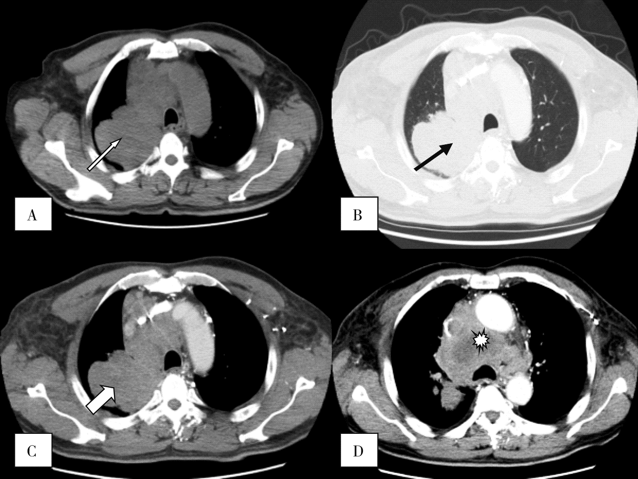
Figure 2
CT findings in a patient with SMARCA4-dNSCLC showing air bronchogram and obstructive changes

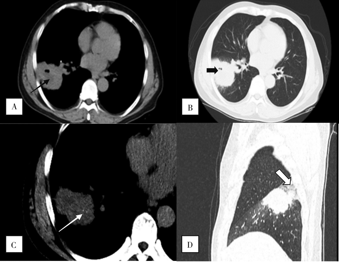
Figure 3
CT findings in a patient with SMARCA4-dNSCLC showing necrosis, vacuoles, and calcification
| [1] | Nicholson AG, Tsao MS, Beasley MB, et al. The 2021 WHO classification of lung tumors: impact of advances since 2015[J]. J Thorac Oncol, 2022, 17(3):362-387. |
| [2] | Crombé A, Alberti N, Villard N, et al. Imaging features of SMARCA4-deficient thoracic sarcomas: a multi-centric study of 21 patients[J]. Eur Radiol, 2019, 29(9):4730-4741. |
| [3] |
Rekhtman N, Montecalvo J, Chang JC, et al. SMARCA4-deficient thoracic sarcomatoid tumors represent primarily smoking-related undifferentiated carcinomas rather than primary thoracic sarcomas[J]. J Thorac Oncol, 2020, 15(2):231-247.
doi: S1556-0864(19)33643-3 pmid: 31751681 |
| [4] | Yoshida A, Kobayashi E, Kubo T, et al. Clinicopathological and molecular characterization of SMARCA4-deficient thoracic sarcomas with comparison to potentially related entities[J]. Mod Pathol, 2017, 30(6):797-809. |
| [5] |
Parikh SA, French CA, Costello BA, et al. NUT midline carcinoma: an aggressive intrathoracic neoplasm[J]. J Thorac Oncol, 2013, 8(10):1335-1338.
doi: 10.1097/JTO.0b013e3182a00f41 pmid: 24457244 |
| [6] | 朱培培, 李新星, 刘佳涵, 等. SMARCA4缺失性肿瘤的临床病理学特征[J]. 中华病理学杂志, 2022, 51(8):792-798. |
|
Zhu PP, Li XX, Liu JH, et al. Clinicopathological features of SMARCA4-deficient tumors[J]. Chinese Journal of Pathology, 2022, 51(8):792-798.
doi: 10.3760/cma.j.cn112151-20220226-00131 pmid: 35922180 |
|
| [7] |
Wong AK, Shanahan F, Chen Y, et al. BRG1, a component of the SWI-SNF complex, is mutated in multiple human tumor cell lines[J]. Cancer Res, 2000, 60(21):6171-6177.
pmid: 11085541 |
| [8] | Kim JH, Woo JH, Lim CY, et al. SMARCA4-deficient non-small cell lung carcinoma: clinicodemographic, computed tomography, and positron emission tomography-computed tomography features[J]. J Thorac Dis, 2024, 16(3):1753-1764. |
| [9] | Okazaki T, Yokoyama K, Tsuchiya J, et al. SMARCA4-deficient thoracic tumor detected by [18F]FDG PET/CT[J]. Eur J Hybrid Imaging, 2021, 5(1):8. |
| [10] | 曹新娜, 李钊, 王全义. SMARCA4缺失型胸部肿瘤17例临床特征及预后分析[J]. 中华结核和呼吸杂志, 2024, 47(4):325-331. |
|
Cao XN, Li Z, Wang QY. Clinical characteristics and prognosis analysis of 17 cases of SMARCA4-deficient chest tumors[J]. Chinese Journal of Tuberculosis and Respiratory Diseases, 2024, 47(4):325-331.
doi: 10.3760/cma.j.cn112147-20230927-00202 pmid: 38599807 |
|
| [11] |
Carter BW, Glisson BS, Truong MT, et al. Small cell lung carcinoma: staging, imaging, and treatment considerations[J]. Radiographics, 2014, 34(6):1707-1721.
doi: 10.1148/rg.346140178 pmid: 25310425 |
| [1] | Fan Xinggang, Liu Jiaxian, Li Chao, Yang Youdong, Gu Wenting, Jiang Xinyang. Computer Aided Diagnosis for COVID-19 in CT Images Utilizing Transfer Learning and Attention Mechanism [J]. J Shanghai Jiaotong Univ Sci, 2025, 30(3): 566-581. |
| [2] | Sun Wenwu, Zhuang Tiange, Chen Siping. Improvement of Prior Image for Metal Artifact Reduction of Computed Tomography [J]. J Shanghai Jiaotong Univ Sci, 2025, 30(3): 446-454. |
| [3] | CHANG Rui, LI Jiqiang, YANG Yanzhao, CHAI Weimin, YAN Fuhua, DONG Haipeng.. Evaluation value of single-phase images from photon-counting CT-based low-dose pancreatic dynamic volume perfusion scanning for pancreatic cancer imaging [J]. Journal of Diagnostics Concepts & Practice, 2025, 24(02): 155-162. |
| [4] | WANG Mengzhen, BAO Shouyu, LIU Peng, YAN Fuhua, YANG Wenjie. Application of photon-counting CT in cardiovascular diseases [J]. Journal of Diagnostics Concepts & Practice, 2025, 24(02): 125-134. |
| [5] | LI Weixia, YAN Fuhua. Photon-counting CT in liver disease: applications and advances [J]. Journal of Diagnostics Concepts & Practice, 2025, 24(02): 118-124. |
| [6] | HUANG Ruikun, YANG Yanzhao, CHAI Weimin. Advances in application of photon-counting CT for pancreatic imaging [J]. Journal of Diagnostics Concepts & Practice, 2025, 24(02): 111-117. |
| [7] | CHENG Dongfeng, ZHOU Ziyi, XU Rongzhong, FANG Zhihong. Two cases of staged treatment of non-small cell lung cancer with traditional Chinese medicine [J]. Journal of Internal Medicine Concepts & Practice, 2025, 20(01): 30-33. |
| [8] | CHEN Weiwei, JIA Hechen, WANG Guoyong, et a(l. Vascular construction of arteriovenous malformation lesions on the body surface Preoperative evaluation and treatment strategies [J]. Journal of Tissue Engineering and Reconstructive Surgery, 2024, 20(6): 617-. |
| [9] | LI Ying, JIANG Han, WANG Xiaoxue, WEI Haonan. Analysis of chest CT findings, diagnosis, and treatment of mucormycosis infection in 65 hematologic disease patients [J]. Journal of Diagnostics Concepts & Practice, 2024, 23(05): 494-499. |
| [10] | LI Zhuohan, HUANG Xinyun, GUO Rui, LI Biao. 18F-FDG PET/CT in the diagnosis and prognosis evaluation of follicular lymphoma [J]. Journal of Diagnostics Concepts & Practice, 2024, 23(04): 439-444. |
| [11] | GAO Meng, CHAI Weimin, YAN Fuhua. Advance in study on diagnosis of pancreatic cystic tumors on CT/MRI imaging [J]. Journal of Diagnostics Concepts & Practice, 2024, 23(02): 184-191. |
| [12] | LI Ming, CHEN Kemin, PAN Zilai, LUO Yu. Research progress on the value of CT and MRI in predicting hemorrhagic transformation after acute ischemic stroke [J]. Journal of Diagnostics Concepts & Practice, 2024, 23(01): 83-89. |
| [13] | LI Jian1,2(李健),ZHU Ye1 (朱晔),GUAN Tianmin1*(关天民). Numerical Simulation Method of Scoliosis Orthosis Considering Muscle Factor [J]. J Shanghai Jiaotong Univ Sci, 2023, 28(4): 486-. |
| [14] | ZHOU Yilei, ZHANG Miao, GUO Rui, ZHOU Jinxin, LI Biao, LI Xiang. Value of 18F-PSMA PET/MRI for early diagnosis of recurrence and metastasis in prostate cancer patients after radical prostatectomy [J]. Journal of Diagnostics Concepts & Practice, 2023, 22(06): 567-572. |
| [15] | DONG Lai, WANG Wei, WU Jialiang, LIU Yanpu, GUAN Xin, CHEN Kemin. Pulmonary imaging manifestations and related research progress of lymphangioleiomyomatosis [J]. Journal of Diagnostics Concepts & Practice, 2023, 22(05): 501-506. |
| Viewed | ||||||
|
Full text |
|
|||||
|
Abstract |
|
|||||