Journal of Diagnostics Concepts & Practice ›› 2024, Vol. 23 ›› Issue (01): 83-89.doi: 10.16150/j.1671-2870.2024.01.011
• Review articles • Previous Articles Next Articles
LI Ming1, CHEN Kemin2( ), PAN Zilai2, LUO Yu1
Received:2023-01-29
Online:2024-02-25
Published:2024-05-30
Contact:
CHEN Kemin
E-mail:keminchenrj@163.com
CLC Number:
LI Ming, CHEN Kemin, PAN Zilai, LUO Yu. Research progress on the value of CT and MRI in predicting hemorrhagic transformation after acute ischemic stroke[J]. Journal of Diagnostics Concepts & Practice, 2024, 23(01): 83-89.
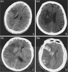
Figure 1
The ECASS classification of HT

| [1] | GBD 2019 Stroke Collaborators. Global, regional, and national burden of stroke and its risk factors, 1990-2019: a systematic analysis for the global burden of disease study[J]. Lancet Neurol, 2021, 20(10):795-820. |
| [2] |
OTT B R, ZMMANI A, KLEEFIELD J, et al. The clinical spectrum of hemorrhagic infarction[J]. Stroke, 1986, 17(4):630-637.
pmid: 3738944 |
| [3] | 《中国脑卒中防治报告2019》编写组. 《中国脑卒中防治报告2019》概要[J]. 中国脑血管病杂志, 2020, 17(5):272-281. |
| Report on Stroke Prevention And Treatment in China Writing Group. Brief report on stroke prevention and treatment in China,2019[J]. Chin J Cerebrovasc Dis, 2020, 17(5): 272-281. | |
| [4] | 中华医学会神经病学分会, 中华医学会神经病学分会脑血管病学组. 中国急性脑梗死后出血转化诊治共识2019[J]. 中华神经科杂志, 2019, 52(4):252-265. |
| Chinese Society of Neurology, Chinese Stroke Society. Consensus on diagnosis and treatment of hemorrhagic transformation after acute ischemic stroke in China 2019[J]. Chin J Neurol, 2019, 52(4):252-265. | |
| [5] |
MEHTA R H, COX M, SMITH E E, et al. Race/ethnic differences in the risk of hemorrhagic complications among patients with ischemic stroke receiving thrombolytic therapy[J]. Stroke, 2014, 45(8):2263-2269.
doi: 10.1161/STROKEAHA.114.005019 pmid: 25070958 |
| [6] | VINAY K, ABUL K A, JON C A, et al. Robbins and Cotran pathologic basis of disease[M]. 9 Ed. Amsterdam: Elsevier, 2015:129-131,1266-1267. |
| [7] |
FRIZZELL J P. Acute stroke: pathophysiology, diagnosis, and Treatment[J]. AACN Clin Issues, 2005, 16(4): 421-440.
doi: 10.1097/00044067-200510000-00002 pmid: 16269890 |
| [8] | HACKE W, KASTE M, FIESCHI C, et al. Randomised double-blind placebo-controlled trial of thrombolytic therapy with intravenous alteplase in acute ischaemic stroke (ECASS II)[J]. Lancet, 1998, 352(9136):1245-1251. |
| [9] | LARRUE V, VON KUMMMER R R, MÜLLER A, et al. Risk for severe hemorrhagic transformation in ischemic stroke patients treated with recombinant tissue plasminogen activator:a secondary analysis of the European-Australasian Acute Stroke Study (ECASS II)[J]. Stroke, 2011, 32(2):438-441. |
| [10] | EMBERSON J, LEES K R, LYDEN P, et al. Effect of treatment delay, age, and stroke severity on the effects of intravenous thrombolysis with alteplase for acute ischae-mic stroke: a meta-analysis of individual patient data from randomised trials[J]. Lancet, 2014, 384(9958):1929-1935. |
| [11] | WHITELEY W N, SLOT K B, FERNANDES P, et al. Risk factors for intracranial hemorrhage in acute ischemic stroke patients treated with recombinant tissue plasminogen activator a systematic review and meta-analysis of 55 studies[J]. Stroke, 2012, 43(11):2904-2909. |
| [12] |
CASTELLANOS M, LEIRA R, SERENA J, et al. Plasma metalloproteinase-9 concentration predicts hemorrhagic transformation in acute ischemic stroke[J]. Stroke, 2003, 34(1):40-46.
pmid: 12511748 |
| [13] |
PRAKKAMAKUL S, YOO A J. ASPECTS CT in acute ischemia: review of current data[J]. Top Magn Reson Imaging, 2017, 26(3):103-112.
doi: 10.1097/RMR.0000000000000122 pmid: 28277460 |
| [14] | DZIALOWSKI I, HILL M D, COUTTS S B, et al. Extent of early ischemic changes on computed tomography (CT) before thrombolysis: prognostic value of the Alberta Stroke Program Early CT Score in ECASS Ⅱ[J]. Stroke, 2006, 37(4):973-978. |
| [15] |
GÁCS G, FOX A J, BARNETT H J, et al. CT visualization of intracranial arterial thromboembolism[J]. Stroke, 1983, 14(5):756-762.
pmid: 6658961 |
| [16] | 黄文磊, 吴雯菁, 姜亦伦. 多模态CT大脑中动脉高密度征对急性缺血性脑卒中出血转化的预测价值的探讨[J]. 医学影像学杂志, 2022, 32(6):919-923. |
| HUANG W L, WU W J, JIANG Y L. Study on the predictive value of high density sign of middle cerebral artery in acute ischemic stroke under multi-modeCT[J]. Med Imaging, 2022, 32(6):919-923. | |
| [17] |
YAGHI S, BOEHME A K, DIBU J, et al. Treatment and outcome of thrombolysis-related hemorrhage: a multicenter retrospective study[J]. JAMA Neurol, 2015, 72(12): 1451-1457.
doi: 10.1001/jamaneurol.2015.2371 pmid: 26501741 |
| [18] | The NINDS t-PA Stroke Study Group. Intracerebral hemorrhage after intravenous t-PA therapy for ischemic stroke[J]. Stroke, 1997, 28(11):2109-2118. |
| [19] |
TIJSSEN M P, HOFMAN P A, STADLER A A, et al. The role of dual energy CT in differentiating between brain haemorrhage and contrast medium after mechanical revascularisation in acute ischaemic stroke[J]. Eur Radiol, 2014, 24(4):834-840.
doi: 10.1007/s00330-013-3073-x pmid: 24258277 |
| [20] | 丁伟莉, 王天宇, 陈青, 等. 急性缺血性卒中血管内介入治疗术后即刻及24h双能CT检查对脑出血转化评估的作用[J]. 中国脑血管病杂志, 2023, 20(4):834-840. |
| DING W L, WANG T Y, CHEN Q, et al. Role of dual-energy CT in different time intervals for the evaluation of postoperative hemorrhagic transformation in endovascular intervention for acute ischemic stroke[J]. Chin J Cerebrovasc Dis, 2023, 20(4):834-840. | |
| [21] |
HORSCH A D, BENNINK E, VAN S T, et al. Computed tomography perfusion derived blood-brain barrier permeability does not yet improve prediction of hemorrhagic transformation[J]. Cerebrovasc Dis, 2018, 45(1-2):26-32.
doi: 10.1159/000485043 pmid: 29402765 |
| [22] |
BANG O Y, GOYAL M, LIEBESKIND D S. Collateral circulation in ischemic stroke: assessment tools and therapeutic strategies[J]. Stroke, 2015, 46(11):3302-3309.
doi: 10.1161/STROKEAHA.115.010508 pmid: 26451027 |
| [23] |
MADELUNG C F, OVESEN C, TRAMPEDACH C, et al. Leptomeningeal collateral status predicts outcome after middle cerebral artery occlusion[J]. Acta Neurol Scand, 2018, 137(1):125-132.
doi: 10.1111/ane.12834 pmid: 28905995 |
| [24] |
LI Q, GAO X, YAO Z, et al. Permeability surface of deep middle cerebral artery territory on computed tomographic perfusion predicts hemorrhagic transformation after stroke[J]. Stroke, 2017, 48(9):2412-2418.
doi: 10.1161/STROKEAHA.117.017486 pmid: 28775139 |
| [25] | BENNINK E, HORSCH A D, DANKBAAR J W, et al. CT perfusion analysis by nonlinear regression for predic-ting hemorrhagic transformation in ischemic stroke[J]. Med Phys, 2015, 42(8):4610-4618. |
| [26] | JAIN A R, JAIN M, KANTHALA A R, et al. Association of CT perfusion parameters with hemorrhagic transformation in acute ischemic stroke[J]. AJNR Am J Neuroradiol, 2013, 34(10):1895-1900. |
| [27] |
BATCHELOR C, PORDELI P, D'ESTERRE C D, et al. Use of noncontrast computed tomography and computed tomographic perfusion in predicting intracerebral hemorrhage after intravenous alteplase therapy[J]. Stroke, 2017, 48(6):1548-1553.
doi: 10.1161/STROKEAHA.117.016616 pmid: 28446625 |
| [28] |
YASSI N, PARSONS M W, CHRISTENSEN S, et al. Prediction of poststroke hemorrhagic transformation using computed tomography perfusion[J]. Stroke, 2013, 44(11):3039-3043.
doi: 10.1161/STROKEAHA.113.002396 pmid: 24003043 |
| [29] |
SOUZA L C, PAYABVASH S, WANG Y, et al. Admission CT perfusion is an independent predictor of hemorrhagic transformation in acute stroke with similar accuracy to DWI[J]. Cerebrovasc Dis, 2012, 33(1):8-15.
doi: 10.1159/000331914 pmid: 22143195 |
| [30] | THOMALLA G, CHENG B, EBINGER M, et al. DWI-FLAIR mismatch for the identification of patients with acute ischaemic stroke within 4.5 h of symptom onset (PRE-FLAIR): a multicentre observational study[J]. Lancet Neurol, 2011, 10(11):978-986. |
| [31] |
KUFNER A, GALINOVIC I, BRUNECKER P, et al. Early infarct flair hyperintensity is associated with increased hemorrhagic transformation after thrombolysis[J]. Eur J Neurol, 2013, 20(2):281-285.
doi: 10.1111/j.1468-1331.2012.03841.x pmid: 22900825 |
| [32] | JHA R, BATTEY T W, PHAM L, et al. Fluid-attenuated inversion recovery hyperintensity correlates with MMP -9 level and hemorrhagic transformation in acute ischemic stroke[J]. Stroke, 2014, 45(4):1040-1045. |
| [33] | LÖVBLAD K O, BAIRD A E, SCHLAUG G, et al. Isc-hemic lesion volumes in acute stroke by diffusion-weighted magnetic resonance imaging correlate with clinical outcome[J]. Ann Neurol, 1997, 42(2):164-170. |
| [34] |
SINGER O C, HUMPICH M C, FIEHLER J, et al. Risk for symptomatic intracerebral hemorrhage after thrombolysis assessed by diffusion-weighted magnetic resonance imaging[J]. Ann Neurol, 2008, 63(1):52-60.
pmid: 17880020 |
| [35] | EL NAWAR R, YEUNG J, LABREUCHE J, et al. MRI-based predictors of hemorrhagic transformation in patients with stroke treated by intravenous thrombolysis[J]. Front Neurol, 2019, 10(897):1-8. |
| [36] | OPPENHEIM C, SAMSON Y, DORMONT D, et al. DWI prediction of symptomatic hemorrhagic transformation in acute MCA infarct[J]. J Neuroradiol, 2002, 29(1):6-13. |
| [37] | SHINODA N, HORI S, MIKAMI K, et al. Prediction of hemorrhagic transformation after acute thrombolysis following major artery occlusion using relative ADC ratio: a retrospective study[J]. J Neuroradiol, 2017, 44(6):361-366. |
| [38] |
BEAUCHAMP M H, BEARE R, DITCHFIELD M, et al. Susceptibility weighted imaging and its relationship to outcome after pediatric traumatic brain injury[J]. Cortex, 2013, 49(2):591-598.
doi: 10.1016/j.cortex.2012.08.015 pmid: 23062584 |
| [39] | TERASAWA Y, YAMAMOTO N, MORIGAKI R, et al. Brush sign on 3T T2-weighted MRI as a potential predictor of hemorrhagic transformation after tissue plasminogen activator therapy[J]. Stroke, 2014, 45(1):274-276. |
| [40] |
NAGARAJA N, TASNEEM N, SHABAN A, et al. Cerebral microbleeds are an independent predictor of hemorrhagic transformation following intravenous alteplase administration in acute ischemic stroke[J]. J Stroke Cerebrovasc Dis, 2018, 27(5):1403-1411.
doi: S1052-3057(17)30718-8 pmid: 29398533 |
| [41] |
ZAND R, TSIVGOULIS G, SINGH M, et al. Cerebral microbleeds and risk of intracerebral hemorrhage post intravenous thrombolysis[J]. J Stroke Cerebrovasc Dis, 2017, 26(3):538-544.
doi: S1052-3057(16)30592-4 pmid: 28065404 |
| [42] |
HJORT N, WU O, ASHKANIAN M, et al. MRI detection of early blood-brain barrier disruption: parenchymal enhancement predicts focal hemorrhagic transformation after thrombolysis[J]. Stroke, 2008, 39(3):1025-1028.
doi: 10.1161/STROKEAHA.107.497719 pmid: 18258832 |
| [43] | LIU H S, CHUNG H W, CHOU M C, et al. Effects of microvascular permeability changes on contrast-enhanced T1 and pharmacokinetic MR imagings after ischemia[J]. Stroke, 2013, 44(7):1872-1877. |
| [44] |
CAMPBELL B C, CHRISTENSEN S, PARSONS M W, et al. Advanced imaging improves prediction of hemorrhage after stroke thrombolysis[J]. Ann Neurol, 2013, 73(4):510-519.
doi: 10.1002/ana.23837 pmid: 23444008 |
| [45] |
MISHRA N K, CHRISTENSEN S, WOUTERS A, et al. Reperfusion of very low cerebral blood volume lesion predicts parenchymal hematoma after endovascular therapy[J]. Stroke, 2015, 46(5):1245-1249.
doi: 10.1161/STROKEAHA.114.008171 pmid: 25828235 |
| [46] |
KIM J H, BANG O Y, LIEBESKIND D S, et al. Impact of baseline tissue status (diffusion-weighted imaging lesion) versus perfusion status (severity of hypoperfusion) on hemorrhagic transformation[J]. Stroke, 2010, 41(3):135-142.
doi: 10.1161/STROKEAHA.109.563122 pmid: 20075362 |
| [47] | LI M, LV Y, WANG M, et al. Magnetic resonance perfusion-weighted imaging in predicting hemorrhagic transformation of acute ischemic stroke:a retrospective study[J]. Diagnostics, 2023, 13(22):3404. |
| [48] | VYAS D, BOHRA V, KARAN V, et al. Rapid processing of perfusion and diffusion for ischemic strokes in the extended time window: an indian experience[J]. Ann Indian Acad Neurol, 2019, 22(1):96-99. |
| [49] | CAMPBELL B C V, MAJOIE C B L M, ALBERS G W, et al. Penumbral imaging and functional outcome in patients with anterior circulation ischaemic stroke treated with endovascular thrombectomy versus medical therapy: a meta-analysis of individual patient-level data[J]. Lancet Neurol, 2019, 18(3):e2. |
| [50] | 曹阳, 刘厚军, 程鸣, 等. MR常规序列及弥散加权成像对脑梗死不同发病时期的诊断价值比较[J]. 安徽医学, 2023, 44(6):702-704. |
| CAO Y, LIU HJ, CHENG M, et al. Comparison of diagnostic value of MR conventional sequence and diffusion-weighted imaging in different stages of cerebral infarction[J]. Anhui Med, 2023, 44(6):702-704. | |
| [51] | YAO L, ZHU H, LIW W, et al. Predictive effect of cortical ribbon sign in DWI on prognosis of mechanical thrombectomy in patients with acute ischemic stroke[J]. Chin J Clin Res, 2023, 36(3):343-346. |
| [52] | 庞伟平, 王保爱. 进展性缺血性脑卒中临床诊疗研究进展[J]. 中国临床研究, 2023, 36(3):380-385. |
| PANG WP, WANG BA. Clinical diagnosis and treatment of progressive stroke[J]. Chin J Clin Res, 2023, 36(3):380-385. |
| [1] | DING Jingfeng, AO Weiqun, ZHU Zhen, SUN Jing, XU Lianggen, ZHENG Shibao, YU Jingjing, HU Jinwen. The value of radiomics based on T2WI and DWI of MRI in preoperative prediction of extramural vascular invasion in rectal cancer [J]. Journal of Diagnostics Concepts & Practice, 2024, 23(01): 46-56. |
| [2] | ZHOU Yilei, ZHANG Miao, GUO Rui, ZHOU Jinxin, LI Biao, LI Xiang. Value of 18F-PSMA PET/MRI for early diagnosis of recurrence and metastasis in prostate cancer patients after radical prostatectomy [J]. Journal of Diagnostics Concepts & Practice, 2023, 22(06): 567-572. |
| [3] | FENG Li, REN Gang, CAI Rong, WANG Xinyun, WANG Hui, ZHU Mingjie. Clinical features study of perivascular epithelioid cell tumor (PEComa) in genitourinary system [J]. Journal of Diagnostics Concepts & Practice, 2023, 22(05): 460-465. |
| [4] | DONG Lai, WANG Wei, WU Jialiang, LIU Yanpu, GUAN Xin, CHEN Kemin. Pulmonary imaging manifestations and related research progress of lymphangioleiomyomatosis [J]. Journal of Diagnostics Concepts & Practice, 2023, 22(05): 501-506. |
| [5] | LI Xiaoshi, QIN Yue. Multiple radiology imaging techniques in the diagnosis of gout [J]. Journal of Diagnostics Concepts & Practice, 2023, 22(03): 311-318. |
| [6] | CHEN Qian, LIN Huimin, YAN Fuhua. Advances in the evaluation of hepatic function by magnetic resonance imaging [J]. Journal of Diagnostics Concepts & Practice, 2023, 22(02): 190-196. |
| [7] | YANG Wenjie, YAN Fuhua. Interpretation of the Clinical Practice Guidelines for Lung Cancer Screening (version 2) of 2022 National Comprehensive Cancer Network(NCCN) [J]. Journal of Diagnostics Concepts & Practice, 2023, 22(01): 14-20. |
| [8] | HUANG Juan, ZHU Xiaolei, LI Xiao, CHEN Kemin, YAN Fuhua, XU Xueqin. Study on blood oxygen level-dependent magnetic resonance imaging for the assessment of early renal hypoxia in chronic kidney disease [J]. Journal of Diagnostics Concepts & Practice, 2022, 21(03): 385-389. |
| [9] | ZHU Naiyi, JIANG Yixin, CHAI Li, CHAI Weimin. Diagnostic values of magnetic resonance imaging in mammography detected BI-RADS≥4 category calcifications with negative ultrasound results [J]. Journal of Diagnostics Concepts & Practice, 2021, 20(05): 439-444. |
| [10] | ZHANG Xuekun, LI Yan, YAN Fuhua, ZHAO Hongfei, SONG Qi. Application value of new accelerating technology based on constellation shuttling imaging in brain MRI [J]. Journal of Diagnostics Concepts & Practice, 2021, 20(04): 378-383. |
| [11] | XU Hao, ZHANG Zhi, XIE Xueqian, YANG Wenyi, LIU Shaowen. Comparative study on software DEEPVESSEL FFR and invasive FFR in assessing coronary ischemia [J]. Journal of Diagnostics Concepts & Practice, 2021, 20(04): 384-390. |
| [12] | SUN Tiantian, YE Baoying, YANG Yu, NIU Jianmei. Color Doppler ultrasound and magnetic resonance imaging in prenatal diagnosis of pernicious placenta previa and pernicious placenta previa with placenta accreta: clinic value and analysis of missed diagnosis [J]. Journal of Diagnostics Concepts & Practice, 2021, 20(02): 173-177. |
| [13] | CAO Juntao, HU Ming, QIAN Pingkang, TU Jianchun, ZHANG Huan, SHEN Junkang. Application value of 3.0T MRI 3D-MERGE sequence in evaluating the degree of supraspinatus tendon injury [J]. Journal of Diagnostics Concepts & Practice, 2021, 20(01): 77-81. |
| [14] | CAO Qiqi, QIN Le, ZHOU Huijuan, YANG Zhitao, SU Wenting, YANG Wenjie, CHENG Zenghui, LU Yong, YAN Fuhua, PAN Zilai. CT features of 2019 novel coronavirus pneumonia [J]. Journal of Diagnostics Concepts & Practice, 2020, 19(1): 16-19. |
| [15] | WU Shuang, XIE Qian, GUAN Xueni, ZHANG Sufang, GAO Xinfang, LIANG Zonghui. Perfomence of MRI intravoxel incoherent motion diffusion weighted imaging parameters in diagnosing active Crohn's disease [J]. Journal of Diagnostics Concepts & Practice, 2020, 19(02): 157-161. |
| Viewed | ||||||
|
Full text |
|
|||||
|
Abstract |
|
|||||