诊断学理论与实践 ›› 2019, Vol. 18 ›› Issue (2): 144-148.doi: 10.16150/j.1671-2870.2019.02.005
钟明, 赵峰, 吴衍, 裴文江, 高航, 郭善禹, 戴谦诚, 张伟( )
)
收稿日期:2019-02-01
出版日期:2019-04-25
发布日期:2019-04-25
通讯作者:
张伟
E-mail:weizh1518@hotmail.com
基金资助:
ZHONG Ming, ZHAO Feng, WU Yan, PEI Wenjiang, GAO Hang, GUO Shanyu, DAI Qiancheng, ZHANG Wei()
Received:2019-02-01
Online:2019-04-25
Published:2019-04-25
Contact:
ZHANG Wei
E-mail:weizh1518@hotmail.com
摘要:
目的:通过建立血浆游离DNA中肿瘤高甲基化基因1(hypermethylated in cancer 1, HIC-1)甲基化检测技术,评估其作为体液活检诊断方法在乳腺良恶性疾病鉴别中的价值。方法:收集35份乳腺疾病患者(其25例乳腺癌,10例乳腺良性疾病)及10份健康志愿者的血液样本,分离、提取血浆中的游离DNA。采用亚硫酸氢盐测序(bisulfite sequencing PCR,BSP)法,检测HIC-1基因启动子区域-636~-424 bp中16个CpG位点的甲基化水平。结果:在检测的16个CpG位点中,以-636~-617 bp中的4个位点甲基化最为明显。其中,乳腺癌组的这4个位点平均甲基化率为22.6%,而良性乳腺疾病组为8.5%,健康对照组为8.3%,3组间的差异有统计学意义(P<0.05)。进一步绘制受试者工作特征曲线(receiver operator characteristic curve, ROC曲线)进行分析,发现以这4个位点的平均甲基化率诊断乳腺癌时,其曲线下面积(area under the cure AUC)为0.794,证实其对乳腺癌具有一定的诊断价值。结论:检测血浆游离DNA中抑癌基因HIC-1启动子区甲基化水平,在乳腺癌诊断中有一定参考价值。
中图分类号:
钟明, 赵峰, 吴衍, 裴文江, 高航, 郭善禹, 戴谦诚, 张伟. 血浆游离DNA中抑癌基因肿瘤高甲基化基因1甲基化检测方法的建立及其在乳腺疾病诊断中的意义[J]. 诊断学理论与实践, 2019, 18(2): 144-148.
ZHONG Ming, ZHAO Feng, WU Yan, PEI Wenjiang, GAO Hang, GUO Shanyu, DAI Qiancheng, ZHANG Wei. Establishment of a detection method for tumor suppressor gene HIC-1 methylation in cell-free DNA and its significance in diagnosis of breast diseases[J]. Journal of Diagnostics Concepts & Practice, 2019, 18(2): 144-148.
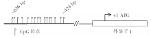
图1
HIC-1启动子甲基化检测模式图及检测区域(NCBI ACCESSION NO. NM_006497.3)
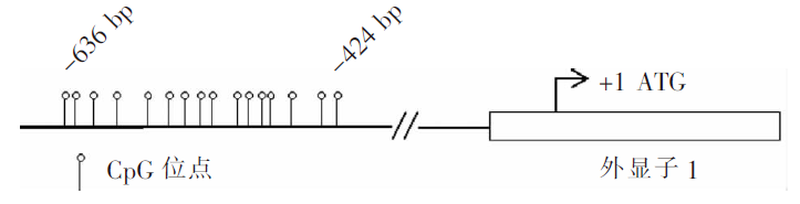

图2
检测区域核苷酸序列(下划线碱基为引物位置)
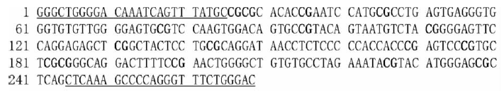
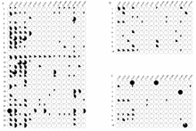
图3
HIC-1启动子区域16个CpG位点甲基化测序结果 A:乳腺癌组;B:良性肿瘤组;C:健康对照组,每行代表1例中10个克隆的测序结果整合,每个圆点代表1个CpG位点,空白代表无甲基化,全黑代表10个克隆全部甲基化,部分黑色代表10个克隆中的甲基化占比情况


图4
血浆HIC-1启动子区域甲基化水平诊断乳腺癌的ROC曲线
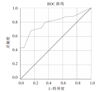
表1
乳腺癌患者血浆HIC-1启动子甲基化水平与其临床病理学间的关系
| 指标 | 例数(n) | 甲基化频率(%) | P值 |
|---|---|---|---|
| 年龄(岁) ≥50 <50 | 18 | 23±14 22±16 | 0.961 |
| 肿瘤直径 ≥2 cm <2 cm | 15 10 | 21±15 24±14 | 0.610 |
| 临床分期 Ⅰ+Ⅱ Ⅲ+Ⅳ | 22 3 | 22±13 23±25 | 0.921 |
| 腋窝淋巴结 阳性 阴性 | 5 20 | 27±19 21±13 | 0.419 |
| ER 阳性 阴性 | 19 6 | 21±14 26±17 | 0.476 |
| PR 阳性 阴性 | 18 7 | 21±14 26±16 | 0.499 |
| HER2 阳性 阴性 | 11 14 | 18±13 26±14 | 0.155 |
| [1] | Allemani C, Matsuda T, di Carlo V, et al. Global surveillance of trends in cancer survival 2000- 14(CONCORD-3): analysis of individual records for 37 513 025 patients diagnosed with one of 18 cancers from 322 population-based registries in 71 countries[J]. Lancet, 2018, 391(10125):1023-1075. |
| [2] | Bae YK, Shim YR, Choi JH, et al. Gene promoter hypermethylation in tumors and plasma of breast cancer patients[J]. Cancer Res Treat, 2005, 37(4):233-240. |
| [3] | Agostini M, Enzo MV, Bedin C, et al. Circulating cell-free DNA: a promising marker of regional lymphonode metastasis in breast cancer patients[J]. Cancer Biomark, 2012, 11(2-3):89-98. |
| [4] | Han ZH, Xu CS, Han H, et al. Value of the level of methylation of RASSF1A and WIF-1 in tissue and serum in neoadjuvant chemotherapeutic assessment for advanced breast cancer[J]. Oncol Lett, 2017, 14(4):4499-4504. |
| [5] | Fu D, Ren C, Tan H, et al. Sox 17 promoter methylation in plasma DNA is associated with poor survival and can be used as a prognostic factor in breast cancer[J]. Medicine (Baltimore), 2015, 94(11):e637 |
| [6] | Parrella P, Scintu M, Prencipe M, et al. HIC 1 promoter methylation and 17p13.3 allelic loss in invasive ductal carcinoma of the breast[J]. Cancer Lett, 2005, 222(1):75-81. |
| [7] | Yamanaka M, Watanabe M, Yamada Y, et al. Altered methylation of multiple genes in carcinogenesis of the prostate[J]. Int J Cancer, 2003, 106(3):382-387. |
| [8] | Kanai Y, Ushijima S, Ochiai A, et al. DNA hypermethylation at the D17S5 locus is associated with gastric carcinogenesis[J]. Cancer Lett, 1998, 122(1-2):135-141. |
| [9] | 裴文江, 赵峰, 吴衍, 等. 肿瘤高甲基化基因1重要下游基因HMMR对乳腺癌细胞株MDA-MB-231迁移和侵袭能力影响的体外实验[J]. 诊断学理论与实践, 2018, 17(1):70-75. |
| [10] | Rasti M, Tavasoli P, Monabati A, et al. Association between HIC1 and RASSF1A promoter hypermethylation with MTHFD1 G1958A polymorphism and clinicopathological features of breast cancer in Iranian patients[J]. Iran Biomed J, 2009, 13(4):199-206. |
| [11] | Rathi A, Virmani AK, Harada K, et al. Aberrant methylation of the HIC1 promoter is a frequent event in speci-fic pediatric neoplasms[J]. Clin Cancer Res, 2003, 9(10 Pt 1):3674-3678. |
| [12] | Narayan G, Arias-Pulido H, Koul S, et al. Frequent promoter methylation of CDH1, DAPK, RARB, and HIC1 genes in carcinoma of cervix uteri: its relationship to clinical outcome[J]. Mol Cancer, 2003, 2:24. |
| [13] | Garcia JM, Silva JM, Dominguez G, et al. Heterogeneous tumor clones as an explanation of discordance between plasma DNA and tumor DNA alterations[J]. Genes Chromosomes Cancer, 2001, 31(3):300-301. |
| [14] | Zhang W, Zeng X, Briggs KJ, et al. A potential tumor suppressor role for Hic1 in breast cancer through transcriptional repression of ephrin-A1[J]. Oncogene, 2010, 29(17):2467-2476. |
| [15] | Wang Y, Weng X, Wang L, et al. HIC1 deletion promotes breast cancer progression by activating tumor cell/fibroblast crosstalk[J]. J Clin Invest, 2018, 128(12):5235-5250. |
| [16] | Skvortsova TE, Rykova EY, Tamkovich SN, et al. Cell-free and cell-bound circulating DNA in breast tumours: DNA quantification and analysis of tumour-related gene methylation[J]. Br J Cancer, 2006, 94(10):1492-1495. |
| [17] | Hernández HG, Tse MY, Pang SC, et al. Optimizing methodologies for PCR-based DNA methylation analysis[J]. Biotechniques, 2013, 55(4):181-197. |
| [18] | 陈卫中, 倪宗瓒, 潘晓平, 等. 用ROC曲线确定最佳临界点和可疑值范围[J]. 现代预防医学, 2005, 32(7):729-731. |
| [19] | Kumar R, Indrayan A. Receiver operating characteristic (ROC) curve for medical researchers[J]. Indian Pediatr, 2011, 48(4):277-287. |
| [1] | 侯筱飒, 杨振江. 前哨淋巴结阳性乳腺癌患者发生非前哨淋巴结转移的危险因素分析[J]. 诊断学理论与实践, 2021, 20(03): 284-289. |
| [2] | 陶志远, 史春颖, 张琦, 宋富贵, 吕哲昊. 数字乳腺体层合成在女性青年期乳腺癌筛查中的应用及研究进展[J]. 诊断学理论与实践, 2021, 20(03): 294-297. |
| [3] | 卢叶君, 陈卉, 张剑, 徐斌, 王冲, 贺烨. 超微血管成像、超声弹性成像联合高频超声在微小乳腺癌中的诊断价值及相关高危超声特征的筛选[J]. 诊断学理论与实践, 2020, 19(04): 391-396. |
| [4] | 罗婷, 周建桥. 超声新技术在预测乳腺癌分子标志物中的应用进展[J]. 诊断学理论与实践, 2020, 19(03): 325-328. |
| [5] | 王志威, 张晓晓, 王杰, 魏敏, 邵玉国, 籍敏, 杨莉, 何奇. 局部晚期乳腺癌患者腋窝淋巴结转移范围的影响因素分析[J]. 诊断学理论与实践, 2019, 18(2): 189-192. |
| [6] | 房莹, 吴东, 常春康. 癌基因与抑癌基因在伯基特淋巴瘤发生发展中的研究进展[J]. 诊断学理论与实践, 2019, 18(06): 630-633. |
| [7] | 裴文江, 赵峰, 吴衍, 钟明, 高航, 李幼生, 顾岩, 郭善禹, 戴谦诚, 张伟. 肿瘤高甲基化基因1重要下游基因HMMR对乳腺癌细胞株MDA-MB-231迁移和侵袭能力影响的体外实验[J]. 诊断学理论与实践, 2018, 17(01): 70-75. |
| [8] | 许海敏, 张培培. 三款自动免疫组织化学染色仪在乳腺癌病理诊断中的应用比较[J]. 诊断学理论与实践, 2017, 16(06): 645-649. |
| [9] | 吴衍, 丁佩芬, 顾岩, 郭善禹, 戴谦诚, 张伟. 乳腺癌肿瘤高甲基化基因1蛋白和透明质酸介导的细胞游走受体表达分析研究[J]. 诊断学理论与实践, 2017, 16(01): 73-78. |
| [10] | 李畅, 方旭前, 卢煌莹, 顾志冬,. QKI-5在三阴乳腺癌组织中的表达及其意义[J]. 诊断学理论与实践, 2016, 15(02): 160-164. |
| [11] | 高玉霞, 沈媛,. P53及nm23在不同分子亚型浸润性乳腺癌中的表达及意义[J]. 诊断学理论与实践, 2015, 14(01): 58-61. |
| [12] | 褚庆华, 张婕, 孙奋勇,. miR-203调控NEDD9抑制乳腺癌细胞MDA-MB-231侵袭及迁移作用机制研究[J]. 诊断学理论与实践, 2014, 13(05): 505-510. |
| [13] | 徐北惠, 梁璆荔, 倪培华,. 乳腺癌细胞自噬与相关miRNA研究进展[J]. 诊断学理论与实践, 2014, 13(04): 429-432. |
| [14] | 曹静, 吕志排, 雷冬梅, 楚天骄, 郝志伟,. 乳腺癌的分子分型与临床病理特征及预后的关系[J]. 诊断学理论与实践, 2013, 12(04): 466-469. |
| [15] | 张俊, 瞿晴, 费晓春, 陈小松, 任若冰, 徐昊平, 许赪, 沈坤炜,. 乳腺癌转移病灶再活检对于指导治疗的价值[J]. 诊断学理论与实践, 2013, 12(03): 304-308. |
| 阅读次数 | ||||||
|
全文 |
|
|||||
|
摘要 |
|
|||||