诊断学理论与实践 ›› 2023, Vol. 22 ›› Issue (06): 567-572.doi: 10.16150/j.1671-2870.2023.06.009
周熠磊, 张淼, 郭睿, 周金鑫, 李彪, 李翔( )
)
收稿日期:2023-05-23
出版日期:2023-12-25
发布日期:2024-03-18
通讯作者:
李翔 E-mail:lx40768@rjh.com.cn基金资助:
ZHOU Yilei, ZHANG Miao, GUO Rui, ZHOU Jinxin, LI Biao, LI Xiang()
Received:2023-05-23
Published:2023-12-25
Online:2024-03-18
摘要:
目的: 探讨18氟-前列腺特异性膜抗原(18F-prostate-specific membrane antigen, 18F-PSMA)-1007正电子发射计算机断层显像/磁共振成像系统(positron emission tomography/magnetic resonance imaging, PET/MRI)(以下简称PET/MRI)检查在早期诊断前列腺癌根治术后患者复发、转移中的价值。方法: 连续纳入2019年6月至2022年1月上海交通大学医学院附属瑞金医院收治的前列腺癌根治术后患者143例,均于术后12~60个月内完成PET/MRI检查。统计PET/MRI检查对患者术后肿瘤复发或转移灶的检出率,根据患者的血清前列腺特异性抗原(prostate-specific antigen, PSA)水平分为PSA≤0.2 ng/mL、0.2 ng/mL<PSA≤0.5 ng/mL、0.5 ng/mL<PSA≤4.0 ng/mL、4.0 ng/mL<PSA<10.0 ng/mL和PSA≥10.0 ng/mL 5组,比较不同组中复发或转移灶的检出率。结果: 25%PSA≤0.2 ng/mL、0.2 ng/mL<PSA≤0.5 ng/mL、0.5 ng/mL<PSA≤4.0 ng/mL、4.0 ng/mL<PSA<10.0 ng/mL和PSA≥10.0 ng/mL组中PET/MRI对复发或转移灶的检出率分别为25.00%(6/24)、70.00%(7/10)、66.67%(16/24)、74.07%(20/27)和94.83%(55/58),各组间两两比较检出率差异有统计学意义(P<0.01或P<0.05)。34例(27.64%)患者根据PET/MRI检查结果改变了治疗方案。结论: PET/MRI在PSA未升高的前列腺癌患者中检出了约1/4存在肿瘤复发或转移的患者,且随着血清PSA水平升高,检出率随之升高,提示PET/MRI可早期检出前列腺癌复发、转移灶,有效指导治疗方案的制定。
中图分类号:
周熠磊, 张淼, 郭睿, 周金鑫, 李彪, 李翔. 18F-PSMA PET/MRI在早期诊断前列腺癌根治术后复发、转移中的价值[J]. 诊断学理论与实践, 2023, 22(06): 567-572.
ZHOU Yilei, ZHANG Miao, GUO Rui, ZHOU Jinxin, LI Biao, LI Xiang. Value of 18F-PSMA PET/MRI for early diagnosis of recurrence and metastasis in prostate cancer patients after radical prostatectomy[J]. Journal of Diagnostics Concepts & Practice, 2023, 22(06): 567-572.
表1
临床资料汇总 (N=143)
| Characteristics | Number of cases |
|---|---|
| Age(year) | |
| Mean age | 67.67 |
| Median age | 68 |
| Range | 43-89 |
| tPSA(ng/mL) | |
| Average value | 46.16 |
| Median | 6 |
| Range | 0.008-1 574.0 |
| Metastasis or recurrence SUVmax | |
| Average value | 24.722 |
| Median | 15.9 |
| Range | 4.6-124.1 |
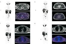
图1
18F-PSMA-1007 PET/MRI全身图像 A:患者男,68岁,术后5年,近期PSA 94 ng/mL,PSMA PET/MRI上可见前列腺术后,术区见结节样放射性摄取增高灶,SUVmax14.6。B:患者男,74岁,术后2年,近期PSA 9.4 ng/mL。右侧髂血管旁可见淋巴结一枚,PSMA-PET放射性摄取明显增高, SUVmax55.4。C:患者男,67岁,术后1年,近期PSA 5.5ng/mL,PSMA PET/MR上可见T6锥体和右侧肩胛骨见斑片及斑点样DWI高信号灶,伴局限性放射性摄取增高,SUVmax27.8。D:患者男,89岁,术后3年,近期PSA 0.15 ng/mL,PSMA PET/MR可见双肺多发斑点状高代谢灶,SUVmax4.83。
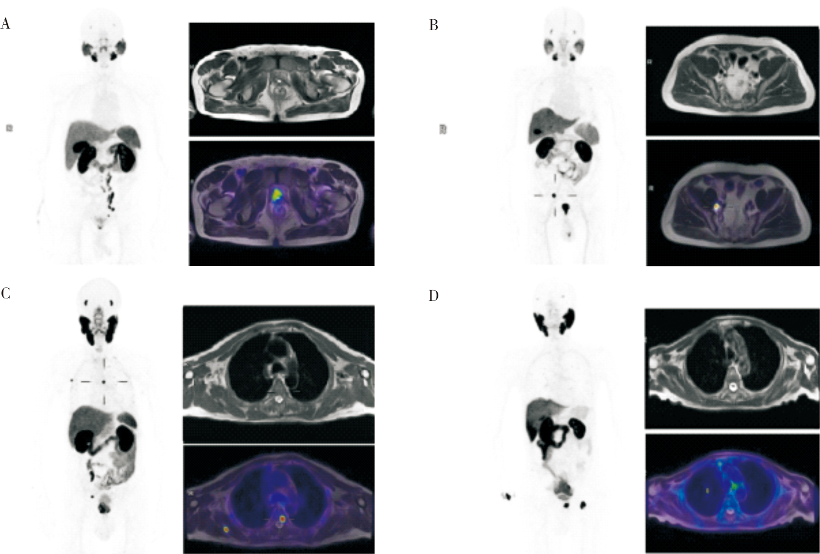
表2
不同PSA水平(ng/mL)分组间PET/MRI对复发或转移灶的检出率比较
| Group | Total number(n) | Number of positive(n) | Positive rate |
|---|---|---|---|
| PSA≤0.2 | 24 | 6 | 25.00% |
| 0.2<PSA≤0.5 | 10 | 7 | 70.00% |
| 0.5<PSA≤4 | 24 | 16 | 66.67% |
| 4<PSA<10 | 27 | 20 | 74.07% |
| PSA≥10 | 58 | 55 | 94.83% |
| [1] |
CHEN W, ZHENG R, BAADE P D, et al. Cancer statistics in China, 2015[J]. CA Cancer J Clin, 2016, 66(2):115-132.
doi: 10.3322/caac.v66.2 URL |
| [2] |
HEIDENREICH A, BASTIAN P J, BELLMUNT J, et al. EAU guidelines on prostate cancer. part 1: screening, diagnosis, and local treatment with curative intent-update 2013[J]. Eur Urol, 2014, 65(1):124-137.
doi: 10.1016/j.eururo.2013.09.046 pmid: 24207135 |
| [3] | 李强. 18F-PSMA-PET/CT在前列腺癌临床诊治中的应用价值探讨[D]. 辽宁: 大连医科大学, 2020. |
| LI Q. Application value of petct in diagnosis and treatment of prostate cancer[D]. Liao Ning: Da Lian Medical University, 2020. | |
| [4] | 沈新平, 叶炯贤, 张娜, 等. 扩散加权成像对诊断前列腺癌的初步研究[J]. 中国医学影像学杂志, 2012, 20(7):485-488. |
| SHEN X P, YE J X, ZHANG N, et al. Preliminary Research for the Diagnosis of Prostate Cancer by Using Diffusion-weighted Imaging[J]. Chin J Med Imaging, 2012, 20(7):485-488. | |
| [5] |
HÖVELS A M, HEESAKKERS R A, ADANG E M, et al. The diagnostic accuracy of CT and MRI in the staging of pelvic lymph nodes in patients with prostate cancer: a meta-analysis[J]. Clin Radiol, 2008, 63(4):387-395.
doi: 10.1016/j.crad.2007.05.022 pmid: 18325358 |
| [6] |
ROWE S P, MACURA K J, CIARALLO A, et al. Comparison of Prostate-Specific Membrane Antigen-Based 18F-DCFBC PET/CT to Conventional Imaging Modalities for Detection of Hormone-Naive and Castration-Resistant Metastatic Prostate Cancer[J]. J Nucl Med, 2016, 57(1):46-53.
doi: 10.2967/jnumed.115.163782 URL |
| [7] |
HOFFMANN M A, MIEDERER M, WIELER H J, et al. Diagnostic performance of 68Gallium-PSMA-11 PET/CT to detect significant prostate cancer and comparison with 18FEC PET/CT[J]. Oncotarget, 2017, 8(67):111073-111083.
doi: 10.18632/oncotarget.v8i67 URL |
| [8] |
PASCHALIS A, SHEEHAN B, RIISNAES R, et al. Prostate-specific Membrane Antigen Heterogeneity and DNA Repair Defects in Prostate Cancer[J]. Eur Urol, 2019, 76(4):469-478.
doi: S0302-2838(19)30520-2 pmid: 31345636 |
| [9] | 陈肖玥, 戴军, 程然, 等. 18F-PSMA联合18F-FDG双核素PET/MR显像对前列腺癌术前评估的临床价值[J]. 中国临床医学影像杂志, 2021, 32(12):876-883. |
| CHEN X Y, DAI J, CHEN R, et al. Clinical value of 18F-PSMA and 18F-FDG dual-nuclide PET/MR imaging in preoperative evaluation of prostate cancer[J]. J Chin Clin Med Imaging, 2021, 32(12):876-883. | |
| [10] | 陈曙光, 胡鹏程, 樊卫, 等. PET/MR全身显像工作流及协议规划专家共识[J]. 中国临床医学, 2020, 27(4):713-721. |
| CHEN S G, HU P C, FAN W, et al. Expert consensus on PET/MR whole body imaging workflow and protocol planning[J]. Chin J Clin Med, 2020, 27(4):713-721. | |
| [11] |
MCGUIRE S. World Cancer Report 2014. Geneva, Switzerland: World Health Organization, International Agency for Research on Cancer, WHO Press, 2015[J]. Adv Nutr, 2016, 7(2):418-419.
doi: 10.3945/an.116.012211 pmid: 26980827 |
| [12] | 龚杨明, 彭鹏, 吴春晓, 等. 2016年上海市前列腺癌发病和死亡情况与2002—2016年变化趋势分析[J]. 肿瘤, 2023, 43(4):297-306. |
|
GONG YM, PENG P, WU CX, et al. Analysis on prostate cancer incidence and mortality in Shanghai 2016 and trends of 2002-2016[J]. Tumor, 2023, 43(04): 297-306.
doi: 10.3781/j.issn.1000-7431.2023.2303-0126 |
|
| [13] | 吉进. 循环外泌体mRNA在前列腺癌早期诊断中作用的研究[D]. 中国人民解放军海军军医大学, 2019. |
| JI J. The role of circulating exosome mRNA in the early diagnosis of prostate cancer[D]. PLA Navy Med Univ, 2019. | |
| [14] | 李曾, 吴毅, 程祝忠, 等. 18F-PSMA-1007PET/CT对前列腺癌根治术后生化复发患者早期诊断评估和临床治疗决策影响的价值研究[J]. 中国癌症杂志, 2021, 31(11):1081-1087. |
| LI Z, WU Y, CHEN Z Z, et al. The value of 18F-PSMA-1007 PET/CT in the early diagnosis and clinical treatment of patients with biochemical recurrence after radical prostatectomy[J]. Chin Oncol, 2021, 31(11):1081-1087 | |
| [15] | 张鑫, 任宏伟, 徐良洲, 等. IVIM-DWI对前列腺癌盆腔淋巴结转移瘤的诊断价值[J]. 临床放射学杂志, 2023, 42(7):1178-1181. |
| Zhang X, Ren H W, XU L Z, et al. The Value of Evaluation Pelvic Lymph Node Metastasis in Prostate Cancer with Intravoxel Incoherent Motion(IVIM) Diffusion Weighted Imaging[J]. J Clin Radiol, 2023, 42(7):1178-1181. | |
| [16] | 周小忠, 黄昌华, 刘德樟, 等. 多模态磁共振成像技术联合前列腺特异性抗原对早期前列腺癌的临床诊断价值[J]. 实用医学影像杂志, 2019, 20(2):197-199. |
| ZHOU X Z, HUANG C H, LIU D Z, et al. Clinical diagnostic value of multimodal magnetic resonance imaging combined with prostate-specific antigen in early prostate cancer[J]. J Pract Med Imaging, 2019, 20(2):197-199. | |
| [17] | JADVAR H. Imaging evaluation of prostate cancer with 18F-fluorodeoxyglucose PET/CT: utility and limitations[J]. Eur J Nucl Med Mol Imaging, 2013, 40 Suppl 1(01):S5-10. |
| [18] | 马兰, 杨吉刚. 前列腺癌PET分子影像学[J]. 临床和实验医学杂志, 2014, 13(7):597-599. |
| MA L, YANG J G. PET molecular imaging of prostate cancer[J]. J Clin Exp Med, 2014, 13(7):597-599. | |
| [19] |
SILVER D A, PELLICER I, FAIR W R, et al. Prostate-specific membrane antigen expression in normal and malignant human tissues[J]. Clin Cancer Res, 1997, 3(1):81-85.
pmid: 9815541 |
| [20] |
MANNWEILER S, AMERSDORFER P, TRAJANOSKI S, et al. Heterogeneity of prostate-specific membrane antigen (PSMA) expression in prostate carcinoma with distant metastasis[J]. Pathol Oncol Res, 2009, 15(2):167-172.
doi: 10.1007/s12253-008-9104-2 pmid: 18802790 |
| [21] |
BOSTWICK D G, PACELLI A, BLUTE M, et al. Prostate specific membrane antigen expression in prostatic intraepithelial neoplasia and adenocarcinoma: a study of 184 cases[J]. Cancer, 1998, 82(11): 2256-2261.
doi: 10.1002/(sici)1097-0142(19980601)82:11<2256::aid-cncr22>3.0.co;2-s pmid: 9610707 |
| [22] |
TROYER J K, BECKETT M L, WRIGHT G L JR. Detection and characterization of the prostate-specific membrane antigen (PSMA) in tissue extracts and body fluids[J]. Int J Cancer, 1995, 62(5):552-558.
doi: 10.1002/ijc.2910620511 pmid: 7665226 |
| [23] |
AFSHAR-OROMIEH A, HETZHEIM H, KRATOCHWIL C, et al. The Theranostic PSMA Ligand PSMA-617 in the Diagnosis of Prostate Cancer by PET/CT: Biodistribution in Humans, Radiation Dosimetry, and First Evaluation of Tumor Lesions[J]. J Nucl Med, 2015, 56(11):1697-1705.
doi: 10.2967/jnumed.115.161299 URL |
| [24] |
SHAMNI O, NEBELING B, GRIEVINK H, et al. Fine-tuning of the automated [18F]PSMA-1007 radiosynthesis[J]. J Labelled Comp Radiopharm, 2019, 62(6):252-258.
doi: 10.1002/jlcr.v62.6 URL |
| [1] | 冯丽, 任刚, 蔡嵘, 汪心韵, 王辉, 祝明洁. 泌尿生殖系统血管周上皮样细胞瘤(PEComa)的临床特征分析[J]. 诊断学理论与实践, 2023, 22(05): 460-465. |
| [2] | 李笑石, 秦越. 影像学技术在痛风诊断及疾病监测中的应用研究进展[J]. 诊断学理论与实践, 2023, 22(03): 311-318. |
| [3] | 李卫侠, 徐学勤, 朱晓雷, 陈克敏. 39例肾上腺皮质癌患者的CT、MRI影像特点及其诊断价值[J]. 诊断学理论与实践, 2023, 22(02): 134-140. |
| [4] | 陈乾, 林慧敏, 严福华. 磁共振成像评估肝功能储备的研究进展[J]. 诊断学理论与实践, 2023, 22(02): 190-196. |
| [5] | 黄娟, 朱晓雷, 李晓, 陈克敏, 严福华, 徐学勤. 血氧水平依赖磁共振成像评估早期慢性肾病肾缺氧的研究[J]. 诊断学理论与实践, 2022, 21(03): 385-389. |
| [6] | 朱乃懿, 姜奕歆, 柴丽, 柴维敏. 磁共振对超声阴性而乳腺X线检出BI-RADS4类以上钙化灶的诊断价值分析[J]. 诊断学理论与实践, 2021, 20(05): 439-444. |
| [7] | 张雪坤, 李彦, 严福华, 赵洪飞, 宋琦. 基于光梭成像的新型加速技术在颅脑MRI中的应用价值研究[J]. 诊断学理论与实践, 2021, 20(04): 378-383. |
| [8] | 孙甜甜, 叶宝英, 杨钰, 牛建梅. 彩色多普勒超声与磁共振成像在凶险型前置胎盘及合并胎盘植入产前诊断中的应用及漏诊分析[J]. 诊断学理论与实践, 2021, 20(02): 173-177. |
| [9] | 于一飞, 王之倩, 李敏, 柳俊, 詹维伟. 自建评分法评估前列腺特异性抗原值在灰区者的前列腺癌风险[J]. 诊断学理论与实践, 2020, 19(03): 314-318. |
| [10] | 吴霜, 解骞, 管雪妮, 张素芳, 高信芳, 梁宗辉. 磁共振体素内不相干运动扩散加权成像诊断活动期克罗恩病的价值及效能分析[J]. 诊断学理论与实践, 2020, 19(02): 157-161. |
| [11] | 曹烨, 刘晓晟, 葛晓乾, 周斌. 运用动态增强磁共振成像评估颈动脉粥样斑块稳定性的初步研究[J]. 诊断学理论与实践, 2019, 18(04): 436-441. |
| [12] | 朱晓雷, 陈璐, 陆文丽, 刘燕, 严福华, 王伟, 董治亚. 474例中枢性性早熟女童不同年龄段垂体MRI影像学异常比例分析[J]. 诊断学理论与实践, 2019, 18(03): 286-290. |
| [13] | 王涛, 邓玉, 赵萍, 于宝华, 王翔, 王朝夫. 基于癌症基因图谱挖掘前列腺癌不同Gleason分级癌组织相关基因分析[J]. 诊断学理论与实践, 2018, 17(06): 694-700. |
| [14] | 李云峰, 江泓, 李宁, 孙青芳. 核磁共振成像诊断三叉神经痛的价值分析与研究[J]. 诊断学理论与实践, 2018, 17(05): 562-565. |
| [15] | 许晶晶, 张敏鸣. 人工智能机器学习方法在阿尔茨海默病中的应用现状[J]. 诊断学理论与实践, 2018, 17(04): 466-470. |
| 阅读次数 | ||||||
|
全文 |
|
|||||
|
摘要 |
|
|||||