诊断学理论与实践 ›› 2024, Vol. 23 ›› Issue (06): 594-601.doi: 10.16150/j.1671-2870.2024.06.006
张慧慧, 方姝, 吴梦雄, 刘方韬, 贺娜英, 董海鹏( ), 严福华
收稿日期:2023-02-22
出版日期:2024-12-25
发布日期:2024-12-25
通讯作者:
董海鹏 E-mail:dhp40427@rjh.com.cn
ZHANG Huihui, FANG Shu, WU Mengxiong, LIU Fangtao, HE Naying, DONG Haipeng(), YAN Fuhua
Received:2023-02-22
Published:2024-12-25
Online:2024-12-25
摘要:
目的: 探讨基于深度学习(deep learning, DL)重建技术的薄层垂体T1WI脂肪抑制(fat suppression, FS)冠状位增强序列,在改善显示垂体神经内分泌肿瘤图像质量中的作用。方法: 前瞻性连续纳入2023年6月至2024年6月诊断或疑似垂体病变患者46例,共有垂体神经内分泌肿瘤病灶40个。所有患者均行薄层垂体DL T1WI FS冠状位增强扫描,并保留未应用DL的原始重建(origin reconstruction, OR)图像,根据重建方式将图像分为DL组和OR组。由2名神经放射诊断医师采用双盲法,分别对2组的图像质量(均匀度、锐利度、伪影、垂体结构辨识度、病灶辨识度、整体质量6个方面)进行主观评估(采用李克特五分量表法),客观评价包括测量并计算垂体神经内分泌肿瘤瘤体和垂体无病灶区的信噪比(signal noise ratio,SNR)和对比噪声比(contrast noise,CNR)。2组图像质量评分差异比较采用Wilcoxon秩和检验,采用组内相关系数ICC(intra-class correlation coefficient,ICC)分别评估2名医师主客观图像测量结果的一致性。结果: DL和OR 2组图像主、客观质量评分的观察者间ICC值均大于0.81,呈极高度一致。在主观图像质量评价方面,DL组和OR组图像的图像均匀度评分分别为4.33(3,5)分、3.73(3,4)分,锐利度评分分别为4.25(3,5)分、3.50(3,4)分,伪影评分分别为4.35(4,5)分、2.95(2,4)分,垂体结构辨识度评分分别为4.38(3,5)分、3.35(2,5)分,病灶辨识度评分分别为4.6(3,5)分、3.15(2,4)分,整体质量评分分别为4.30(4,5)分、2.63(2,3)分,DL组均高于OR组,差异有统计学意义(P均<0.001)。客观图像质量方面,DL组和OR组垂体瘤的SNR分别为26.96(18.10,34.15)、16.51(11.24,20.65),CNR分别为11.30(6.74,19.72)、4.34(3.07,6.00),垂体的SNR分别为38.36(31.93,47.03)、17.02(15.49,20.51),CNR分别29.89(23.28,39.75)、18.44(16.61,24.56),DL组均高于OR组,差异有统计学意义(P均<0.001)。2名医师评估所得的主客观指标一致性较好。结论: 基于DL的T1WI FS冠状位增强序列,在确保图像空间分辨率的情况下,可明显改善垂体神经内分泌肿瘤图像质量,提升图像SNR及CNR,为临床诊疗提供精准的影像学依据。
中图分类号:
张慧慧, 方姝, 吴梦雄, 刘方韬, 贺娜英, 董海鹏, 严福华. 深度学习重建技术在改善磁共振冠状位T1WI显示垂体神经内分泌肿瘤图像质量中的研究[J]. 诊断学理论与实践, 2024, 23(06): 594-601.
ZHANG Huihui, FANG Shu, WU Mengxiong, LIU Fangtao, HE Naying, DONG Haipeng, YAN Fuhua. Study on deep learning reconstruction technology in improving image quality of pituitary neuroendocrine tumors in coronal T1WI magnetic resonance image[J]. Journal of Diagnostics Concepts & Practice, 2024, 23(06): 594-601.
表1
2名医师主观图像质量评价指标的一致性分析结果[ICC值(95%可信区间)]
| Subjective evaluation indicators | DL T1WI FS | OR T1WI FS | |||||
|---|---|---|---|---|---|---|---|
| Doctor 1 | Doctor 2 | ICC | Doctor 1 | Doctor 2 | ICC | ||
| Uniformity | 4.33(3,5) | 4.35(4,5) | 0.92(0.85~0.96) | 3.73(3,4) | 3.78(3,4) | 0.88(0.76~0.93) | |
| Sharpness | 4.25(3,5) | 4.28(3,5) | 0.90(0.81~0.95) | 3.50(3,4) | 3.53(3,4) | 0.86(0.73~0.93) | |
| Artifacts | 4.35(4,5) | 4.38(4,5) | 0.84(0.71~0.91) | 2.95(2,4) | 2.93(2,4) | 0.87(0.75~0.93) | |
| Recognition of pituitary structure | 4.38(3,5) | 4.45(4,5) | 0.93(0.87~0.96) | 3.35(2,5) | 3.38(3,5) | 0.89(0.79~0.94) | |
| Recognition of the lesion | 4.6(3,5) | 4.53(3,5) | 0.85(0.71~0.92) | 3.15(2,4) | 3.18(2,4) | 0.87(0.74~0.93) | |
| Overall quality | 4.30(4,5) | 4.4(4,5) | 0.89(0.79~0.94) | 2.63(2,3) | 2.68(2,3) | 0.88(0.77~0.94) | |
表2
2名医师客观图像质量评价指标的一致性分析结果[ICC值(95%可信区间)]
| Objective evaluation indicators | DL T1WI FS | OR T1WI FS | |||||
|---|---|---|---|---|---|---|---|
| Doctor 1 | Doctor 2 | ICC | Doctor 1 | Doctor 2 | ICC | ||
| SNR adenoma | 26.34(11.42,45.55) | 28(10.45,70.66) | 0.859(0.73~0.93) | 16.76(7.8,30.26) | 17.39(8.06,34.55) | 0.912(0.83~0.95) | |
| CNR adenoma | 13.72(3.56,30.9) | 12.65(0.41,31.81) | 0.949(0.90~0.97) | 4.44(1.12,8.51) | 4.68(1.9,10.49) | 0.889(0.79~0.94) | |
| SNR pituitary | 39.92(18.76,67.79) | 39.72(21.82,75.51) | 0.934(0.88~0.97) | 18.42(11.21,28.43) | 18.2(11.64,26.41) | 0.876(0.77~0.93) | |
| CNR pituitary | 32.12(13.36,63.39) | 30.46(10.45,55.09) | 0.891(0.79~0.94) | 20.73(10.89,42.76) | 21.4(11.61,43.8) | 0.907(0.83~0.95) | |
表3
2组图像主观评价比较结果
| Subjective evaluation indicators | DL T1WI FS | OR T1WI FS | Value of Z | Value of P |
|---|---|---|---|---|
| Uniformity | 4.33(3,5) | 3.73(3,4) | -4.69 | <0.001 |
| Sharpness | 4.25(3,5) | 3.50(3,4) | -5.04 | <0.001 |
| Artifacts | 4.35(4,5) | 2.95(2,4) | -7.96 | <0.001 |
| Recognition of pituitary structure | 4.38(3,5) | 3.35(2,5) | -6.14 | <0.001 |
| Recognition of the lesion | 4.6(3,5) | 3.15(2,4) | -7.25 | <0.001 |
| Overall quality | 4.30(4,5) | 2.63(2,3) | -8.04 | <0.001 |
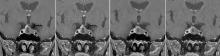
图1
54岁女性垂体大腺瘤患者的T1WI FS冠状位图像 可见鞍隔膨隆,鞍底稍下陷,病灶最大直径18 mm。A、C:DL T1WI FS冠状位图像;B、D:OR T1WI FS冠状位图像。与OR T1WI FS图像相比,DL T1WI FS图像所显示的垂体病灶边界显示更加清晰,图像均匀度、锐利度、垂体结构辨识度、病灶辨识度更好,噪声明显减小。
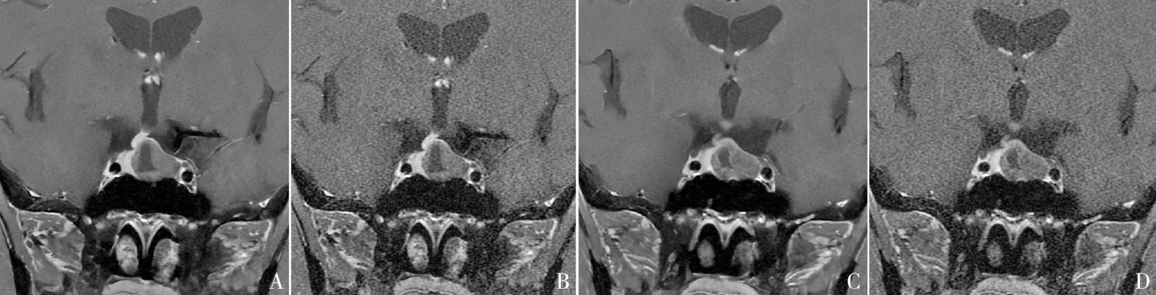

图2
23岁男性垂体大腺瘤患者的T1WI FS 冠状位图像 可见右侧鞍底-海绵窦区结节,垂体柄稍左偏,病灶最大直径12 mm。A、C:DL T1WI FS冠状位图像;B、D:OR T1WI FS冠状位图像。与OR T1WI FS图像相比,DL T1WI FS所显示图像病灶边缘勾勒更为清晰,图像细节显示清晰,均匀度、锐利度、病灶辨识度更好,噪声更小。

图3
38岁男性垂体微腺瘤患者的T1WI FS冠状位图像 可见垂体右翼结节,垂体形态饱满,垂体柄略向左侧偏移,病灶最大直径8 mm。A、C:DL T1WI FS冠状位图像;B、D:OR T1WI FS冠状位图像。与OR T1WI FS图像相比,DL T1WI FS所显示垂体病灶轮廓清晰,图像均匀度、锐利度、垂体结构辨识度、病灶辨识度更好,噪声更小。
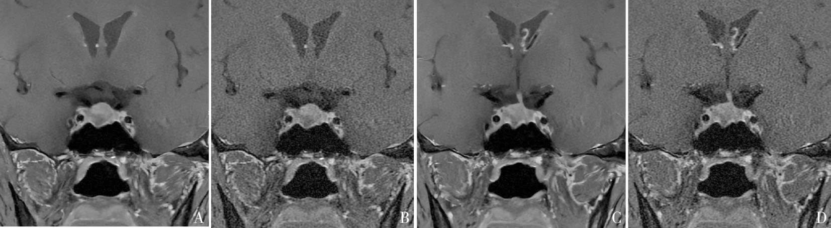
表4
2组图像客观评价比较结果
| Objective evaluation indicators | DL T1WI FS | OR T1WI FS | Value of Z | Value of P |
|---|---|---|---|---|
| SNR adenoma | 26.96(18.10,34.15) | 16.51(11.24,20.65) | -4.44 | <0.000 1 |
| CNR adenoma | 11.30(6.74,19.72) | 4.34(3.07,6.00) | -5.55 | <0.000 1 |
| SNR pituitary | 38.36(31.93,47.03) | 17.02(15.49,20.51) | -5.44 | <0.000 1 |
| CNR pituitary | 29.89(23.28,39.75) | 18.44(16.61,24.56) | -4.27 | <0.000 1 |
| [1] |
DI IEVA A, ROTONDO F, SYRO L V, et al. Aggressive pituitary adenomas--diagnosis and emerging treatments[J]. Nat Rev Endocrinol, 2014, 10(7):423-435.
doi: 10.1038/nrendo.2014.64 pmid: 24821329 |
| [2] |
WANG X, DAI Y, LIN H, et al. Shape and texture analyses based on conventional MRI for the preoperative prediction of the aggressiveness of pituitary adenomas[J]. Eur Radiol, 2023, 33(5):3312-3321.
doi: 10.1007/s00330-023-09412-7 pmid: 36738323 |
| [3] | 林绍坚, 吴隽宸, 吴哲褒. 加强垂体神经内分泌肿瘤的免疫治疗研究[J]. 中华内分泌代谢杂志, 2024, 40(11):920-922. |
| LIN S J, WU J C, WU Z B, et al. Enhancing resreach on immunotherapy for pitutiary neuroendocrine tumors[J]. Chin J Endocrinol Metab, 2024, 40(11): 920-922. | |
| [4] | LEE D H, PARK J E, NAM Y K, et al. Deep learning-based thin-section MRI reconstruction improves tumour detection and delineation in pre- and post-treatment pitui-tary adenoma[J]. Sci Rep, 2021, 11(1):21302. |
| [5] |
TRITOS N A, MILLER K K. Diagnosis and management of pituitary adenomas: A review[J]. JAMA, 2023, 329(16):1386-1398.
doi: 10.1001/jama.2023.5444 pmid: 37097352 |
| [6] | BONNEVILLE J F, POTORAC J, BECKERS A. Neuroimaging of aggressive pituitary tumors[J]. Rev Endocr Metab Disord, 2020, 21(2):235-242. |
| [7] |
KIM M, KIM H S, KIM H J, et al. Thin-slice pituitary MRI with deep learning-based reconstruction: diagnostic performance in a postoperative setting[J]. Radiology, 2021, 298(1):114-122.
doi: 10.1148/radiol.2020200723 pmid: 33141001 |
| [8] | KIM M, KIM H S, PARK J E, et al. Thin-slice pituitary MRI with deep learning-based reconstruction for preo-perative prediction of cavernous sinus invasion by pitui-tary adenoma: a prospective study[J]. AJNR Am J Neuroradiol, 2022, 43(2):280-285. |
| [9] | RASTOGI A, BRUGNARA G, FOLTYN-DUMITRU M, et al. Deep-learning-based reconstruction of under-sampled MRI to reduce scan times: a multicentre, retrospective, cohort study[J]. Lancet Oncol, 2024, 25(3):400-410. |
| [10] | 严福华. 深度学习图像重建算法的临床应用和发展前景[J]. 中华放射学杂志, 2022, 56(11):1165-1167. |
| YAN F H. The clinical application and development prospect of deep learning reconstruction algorithm[J]. Chin J Radiol, 2022, 56(11):1165-1167. | |
| [11] |
VAN DER VELDE N, HASSING H C, BAKKER B J, et al. Improvement of late gadolinium enhancement image quality using a deep learning-based reconstruction algorithm and its influence on myocardial scar quantification[J]. Eur Radiol, 2021, 31(6):3846-3855.
doi: 10.1007/s00330-020-07461-w pmid: 33219845 |
| [12] | DUBLJEVIC N, MOORE S, LAUZON M L, et al. Effect of MR head coil geometry on deep-learning-based MR image reconstruction[J]. Magn Reson Med, 2024, 92(4):1404-1420. |
| [13] | SHIRAISHI K, NAKAURA T, UETANI H, et al. Deep learning-based reconstruction and 3D hybrid profile order technique for MRCP at 3T: evaluation of image qua-lity and acquisition time[J]. Eur Radiol, 2023, 33(11):7585-7594. |
| [14] | ICHINOHE F, OYAMA K, YAMADA A, et al. Usefulness of breath-hold fat-suppressed T2-weighted images with deep learning-based reconstruction of the liver: comparison to conventional free-breathing turbo spin echo[J]. Invest Radiol, 2023, 58(6):373-379. |
| [15] | 方姝, 吴梦雄, 陈乾, 等. 深度学习在肝脏屏气T2加权成像图像质量评价中的应用研究[J]. 磁共振成像, 2023, 14(5):31-35,40. |
| FANG S, WU M X, CHEN Q, et al. Clinical feasibility of breath-hold fat-suppressed T2-weighted sequence with deep learning reconstruction for liver imaging[J]. Chin J Magn Reson Imaging, 2023, 14(5): 31-35+40. | |
| [16] | CHAIKA M, AFAT S, WESSLING D, et al. Deep learning-based super-resolution gradient echo imaging of the pancreas: Improvement of image quality and reduction of acquisition time[J]. Diagn Interv Imaging, 2023, 104(2):53-59. |
| [17] |
ALMANSOUR H, GASSENMAIER S, NICKEL D, et al. Deep learning-based superresolution reconstruction for upper abdominal magnetic resonance imaging: An analysis of image quality, diagnostic confidence, and lesion conspicuity[J]. Invest Radiol, 2021, 56(8):509-516.
doi: 10.1097/RLI.0000000000000769 pmid: 33625063 |
| [18] | SAUER S T, CHRISTNER S A, LOIS A M, et al. Deep learning k-space-to-image reconstruction facilitates high spatial resolution and scan time reduction in diffusion-weighted imaging breast MRI[J]. J Magn Reson Imaging, 2024, 60(3):1190-1200. |
| [19] | LEE K L, KESSLER D A, DEZONIE S, et al. Assessment of deep learning-based reconstruction on T2-weighted and diffusion-weighted prostate MRI image quality[J]. Eur J Radiol, 2023, 166:111017. |
| [20] |
FEUERRIEGEL G C, WEISS K, KRONTHALER S, et al. Evaluation of a deep learning-based reconstruction method for denoising and image enhancement of shoulder MRI in patients with shoulder pain[J]. Eur Radiol, 2023, 33(7):4875-4884.
doi: 10.1007/s00330-023-09472-9 pmid: 36806569 |
| [21] |
ESTLER A, HAUSER T K, BRUNNÉE M, et al. Deep learning-accelerated image reconstruction in back pain-MRI imaging: reduction of acquisition time and improvement of image quality[J]. Radiol Med, 2024, 129(3):478-487.
doi: 10.1007/s11547-024-01787-x pmid: 38349416 |
| [22] | KANIEWSKA M, DEININGER-CZERMAK E, GETZMANN J M, et al. Application of deep learning-based image reconstruction in MR imaging of the shoulder joint to improve image quality and reduce scan time[J]. Eur Radiol, 2023, 33(3):1513-1525. |
| [23] |
YOO H, YOO R E, CHOI S H, et al. Deep learning-based reconstruction for acceleration of lumbar spine MRI: a prospective comparison with standard MRI[J]. Eur Radiol, 2023, 33(12):8656-8668.
doi: 10.1007/s00330-023-09918-0 pmid: 37498386 |
| [24] | XIE Y, LI X, HU Y, et al. Deep learning reconstruction for turbo spin echo to prospectively accelerate ankle MRI: A multi-reader study[J]. Eur J Radiol, 2024, 175:111451. |
| [25] |
SUH P S, PARK J E, ROH Y H, et al. Improving diagnostic performance of MRI for temporal lobe epilepsy with deep learning-based image reconstruction in patients with suspected focal epilepsy[J]. Korean J Radiol, 2024, 25(4):374-383.
doi: 10.3348/kjr.2023.0842 pmid: 38528695 |
| [26] | CHAIKA M, BRENDEL J M, URSPRUNG S, et al. Deep learning reconstruction of prospectively accelerated MRI of the pancreas: clinical evaluation of shortened breath-hold examinations with dixon fat suppression[J/OL]. Invest Radiol, 2024-07-23. https://journals.lww.com/investigativeradiology/abstract/9900/deep_learning_reconstruction_of_prospectively.235.aspx. |
| [27] | SHANBHOGUE K, TONG A, SMEREKA P, et al. Accele-rated single-shot T2-weighted fat-suppressed (FS) MRI of the liver with deep learning-based image reconstruction: qualitative and quantitative comparison of image quality with conventional T2-weighted FS sequence[J]. Eur Radiol, 2021, 31(11):8447-8457. |
| [28] | PARK H, NAM Y K, KIM H S, et al. Deep learning-based image reconstruction improves radiologic evaluation of pituitary axis and cavernous sinus invasion in pitui-tary adenoma[J]. Eur J Radiol, 2023, 158:110647. |
| [29] | GONG K, HAN P, EL FAKHRI G, et al. Arterial spin labeling MR image denoising and reconstruction using unsupervised deep learning[J]. NMR Biomed, 2022, 35(4):e4224. |
| [30] |
KAKIGI T, SAKAMOTO R, TAGAWA H, et al. Diagnostic advantage of thin slice 2D MRI and multiplanar reconstruction of the knee joint using deep learning based denoising approach[J]. Sci Rep, 2022, 12(1):10362.
doi: 10.1038/s41598-022-14190-1 pmid: 35725760 |
| [31] | DALY A F, BECKERS A. The epidemiology of pituitary adenomas[J]. Endocrinol Metab Clin North Am, 2020, 49(3):347-355. |
| [32] | LIU Z, WEN B, WANG Z, et al. Deep learning-based reconstruction enhances image quality and improves diagnosis in magnetic resonance imaging of the shoulder joint[J]. Quant Imaging Med Surg, 2024, 14(4):2840-2856. |
| [33] |
WANG X, MA J, BHOSALE P, et al. Novel deep learning-based noise reduction technique for prostate magnetic resonance imaging[J]. Abdom Radiol (NY), 2021, 46(7):3378-3386.
doi: 10.1007/s00261-021-02964-6 pmid: 33580348 |
| [34] | TAO Q, WANG K, WEN B, et al. Assessment of image quality and diagnostic accuracy for cervical spondylosis using T2W-STIR sequence with a deep learning-based reconstruction approach[J]. Eur Spine J, 2024, 33(8):2982-2996. |
| [1] | 周恒花, 林斓, 朱桂香, 刘敏, 黄文涛. 2例膀胱纯上皮性神经内分泌肿瘤临床病理特征差异及文献复习[J]. 诊断学理论与实践, 2024, 23(06): 602-611. |
| [2] | 查云飞, 武夏夏. MRI深度学习图像重建技术在肌骨系统疾病诊断的应用进展[J]. 诊断学理论与实践, 2024, 23(02): 114-118. |
| [3] | 吕晓宇, 冯威铭, 周慧赟, 李纪强, 董海鹏, 黄娟. 基于磁共振深度学习重建算法缩短扫描时间的可行性分析:水模研究[J]. 诊断学理论与实践, 2024, 23(02): 131-138. |
| [4] | 钱佳乐, 范婧, 朱宏, 王落桐, 孔德艳. 深度学习图像重建在虚拟平扫CT尿路成像中的应用价值[J]. 诊断学理论与实践, 2024, 23(02): 139-145. |
| [5] | 高梦, 柴维敏, 严福华. 胰腺囊性肿瘤的CT/MRI诊断进展[J]. 诊断学理论与实践, 2024, 23(02): 184-191. |
| [6] | 李明, 陈克敏, 潘自来, 罗禹. CT及MRI预测急性缺血性脑梗死出血性转化的价值研究进展[J]. 诊断学理论与实践, 2024, 23(01): 83-89. |
| [7] | 丁景峰, 敖炜群, 朱珍, 孙静, 徐良根, 郑世保, 俞晶晶, 胡金文. 基于T2WI和DWI的磁共振影像组学在术前预测直肠癌壁外血管侵犯的价值研究[J]. 诊断学理论与实践, 2024, 23(01): 46-56. |
| [8] | 周熠磊, 张淼, 郭睿, 周金鑫, 李彪, 李翔. 18F-PSMA PET/MRI在早期诊断前列腺癌根治术后复发、转移中的价值[J]. 诊断学理论与实践, 2023, 22(06): 567-572. |
| [9] | 冯丽, 任刚, 蔡嵘, 汪心韵, 王辉, 祝明洁. 泌尿生殖系统血管周上皮样细胞瘤(PEComa)的临床特征分析[J]. 诊断学理论与实践, 2023, 22(05): 460-465. |
| [10] | 李笑石, 秦越. 影像学技术在痛风诊断及疾病监测中的应用研究进展[J]. 诊断学理论与实践, 2023, 22(03): 311-318. |
| [11] | 李卫侠, 徐学勤, 朱晓雷, 陈克敏. 39例肾上腺皮质癌患者的CT、MRI影像特点及其诊断价值[J]. 诊断学理论与实践, 2023, 22(02): 134-140. |
| [12] | 颜凌, 王凌云, 陈勇, 杜联军. 双能CT图像深度学习重建算法在胃癌术前T分期中的应用[J]. 诊断学理论与实践, 2023, 22(02): 154-159. |
| [13] | 陈乾, 林慧敏, 严福华. 磁共振成像评估肝功能储备的研究进展[J]. 诊断学理论与实践, 2023, 22(02): 190-196. |
| [14] | 张美玲, 朱潇邦, 宋爱玲, 周剑平, 李庆云. 小细胞肺癌患者采用PD-L1抑制剂治疗致垂体炎1例报道并文献复习[J]. 诊断学理论与实践, 2022, 21(06): 741-745. |
| [15] | 范婧, 杨文洁, 王梦真, 陆伟, 石骁萌, 朱宏. 深度学习重建算法在低管电压冠状动脉CT血管成像中的应用[J]. 诊断学理论与实践, 2022, 21(03): 374-379. |
| 阅读次数 | ||||||
|
全文 |
|
|||||
|
摘要 |
|
|||||