诊断学理论与实践 ›› 2025, Vol. 24 ›› Issue (01): 100-105.doi: 10.16150/j.1671-2870.2025.01.015
龚静青a, 曹端荣b, 庄义欣c, 邱立a, 李晓鸣a( )
)
收稿日期:2022-04-12
接受日期:2024-08-30
出版日期:2025-02-25
发布日期:2025-02-25
通讯作者:
李晓鸣 E-mail:2687528433@qq.com
GONG Jingqinga, CAO Duanrongb, ZHUANG Yixinc, QIU Lia, LI Xiaominga()
Received:2022-04-12
Accepted:2024-08-30
Published:2025-02-25
Online:2025-02-25
摘要:
本文报道1例罕见的双表型鼻腔鼻窦肉瘤(biphenotypic sinonasal sarcoma, BSNS)患者,分析其临床病理特征。患者为35岁男性,因“反复右鼻涕中带血3月余”入院。行鼻内镜右侧鼻腔鼻窦颅底肿物切除术,术后病理活检。在光镜下观察,见肿瘤边界欠清晰,表面被覆纤毛柱状上皮细胞,局灶纤毛上皮细胞鳞状化生,黏膜内陷扩张及黏膜腺体增生。肿瘤由弥漫梭形细胞构成,细胞排列较密集,呈束状、编织状或人字形排列,细胞无明显异型性;染色质细腻,未见明显核分裂象和坏死;间质内见薄壁、扩张、分枝状的血管,呈鹿角样,未见明确横纹肌样分化区域。肿瘤组织的免疫表型为,Vimentin、H-Caldesmon、INI-1、Bcl-2和CD99呈阳性表达,Calponin、S100、MyoD1、SMA呈部分阳性表达,Ki-67增殖指数在热点区约为35%+;Myogenin、STAT6、SOX-10、Desmin、β-catenin、CK、CD34、NSE、PR和EMA均为阴性。基因检测显示,PAX-3基因断裂,FISH检测结果为阳性;SYT基因无断裂,FISH检测结果为阴性。该病例临床表现特征缺乏特异性,但被覆纤毛柱状上皮呈腺腔样结构,下陷于梭形细胞内呈囊性扩张,或梭形细胞内见增生的呼吸道腺体,是BSNS其相对典型的组织学特征。BSNS与其他梭型细胞肿瘤鉴别,应结合鼻腔鼻窦的发病部位、肿瘤由相对温和的梭形细胞构成、相对典型的组织学特征、表达肌源性和神经源性的免疫标记及特征性的PAX3基因检测进行综合诊断。
中图分类号:
龚静青, 曹端荣, 庄义欣, 邱立, 李晓鸣. 双表型鼻腔鼻窦肉瘤1例临床病理分析[J]. 诊断学理论与实践, 2025, 24(01): 100-105.
GONG Jingqing, CAO Duanrong, ZHUANG Yixin, QIU Li, LI Xiaoming. Clinicopathological analysis of biphenotypic sinonasal sarcoma: a case report[J]. Journal of Diagnostics Concepts & Practice, 2025, 24(01): 100-105.
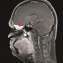
图1
MRI T1增强扫描示右侧中上鼻道及筛窦内见不规则异常信号影,边缘清晰(箭头示)
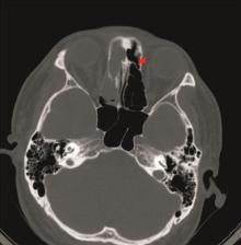
图2
CT检查示筛窦内筛板骨质部分破坏(箭头示)

图3
肿瘤病理镜下特征A:肿瘤边界不清,表面被覆正常纤毛柱状上皮(10×10)。B:肿瘤表面被覆鳞状上皮(5×10)。C:上皮下陷于肿瘤细胞内并囊性扩张(10×10)。D:肿瘤内见增生的呼吸道腺体聚集(20×10)。
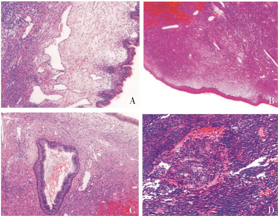
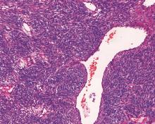
图4
肿瘤细胞呈束状或鱼骨样排列,可见鹿角样血管(20×10)
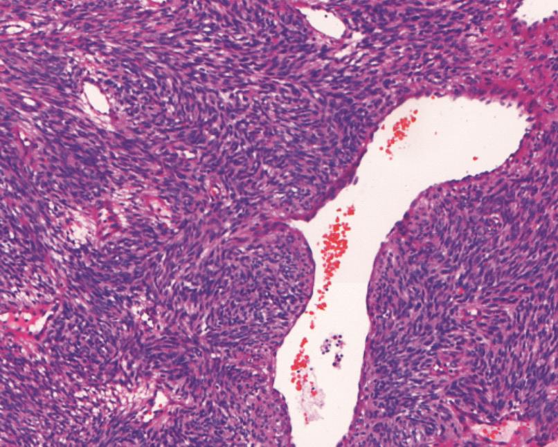

图5
肿瘤侵犯骨组织(10×10)
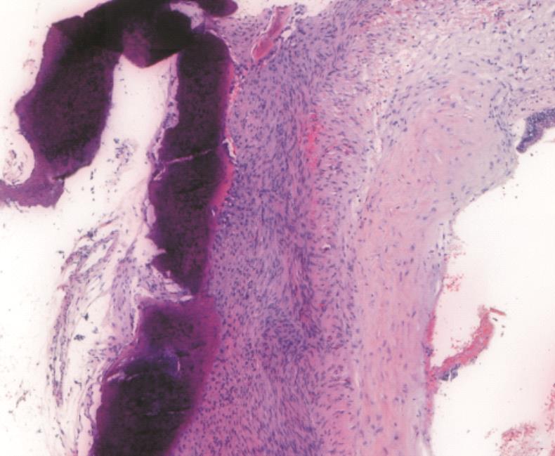
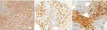
图6
免疫组化结果A:免疫组织化学弥漫表达H-Caldesmon (EnVision法);B:免疫组织化学局灶表达MyoD1蛋白(EnVision法);C:免疫组织化学局灶表达S100蛋白(EnVision法)。

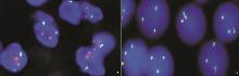
图7
基因检测结果A:PAX3基因检测约76%的肿瘤细胞存在红、绿信号分离,证实存在PAX3有断裂FISH(高倍);B:SYT基因检测肿瘤细胞无红、绿信号分离,证实SYT基因检测无断裂FISH(高倍)。
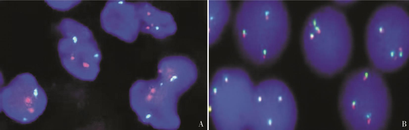
表1
具有高级别转化的BSNS的临床病理学特征
| 年龄/性别 | 72岁/女 | 66岁/男 | 67岁/男 |
|---|---|---|---|
| 发病部位 | 右上颌窦、筛窦 | 左侧眶上肿物 | 右侧筛窦、上颌窦和额窦 |
| 肿瘤侵犯周围组织情况 | 侵犯双侧眶内和颅内 | 侵犯左颅内 | 侵犯鼻外眶 |
| 高级别区域组织学形态 | 高级别梭形细胞肉瘤 | 高级别梭形细胞肉瘤 | 高级别横纹肌肉瘤 |
| 高级别区域免疫组化 | Myogenin、Desmin阳性 | 灶性SMA和S100阳性 | MyoD1、PAX7 Myogenin、Desmin阳性 |
| 分子遗传学 | PAX3基因断裂,9p和22号拷贝数改变 | PAX::MAML3融合 | PAX::MAML3融合 |
| 病程 | 2年鼻塞病史伴间歇性鼻出血、头疼及嗅觉下降 | 15年前切除左鼻腔肿物,当时诊断滑膜肉瘤,现左眼复视及脓性鼻涕 | 3年前接受过鼻中隔形成术、双侧额窦造口和右中鼻甲大块切除术 |
| 治疗及预后 | 手术切除术,术后4.5个月急性冠状动脉疾病,肿物无复发 | 手术治疗,放疗,术后10个月未复发 | 新辅助化疗、手术切除术、放疗,4个月后复发,15个月死亡 |
表2
BSNS免疫组化表型
| 标记物 | 阳性数(例) | 总数(例) | 阳性数/总数 | 百分比(%) |
|---|---|---|---|---|
| S100 | 93 | 94 | 93/94 | 98.93 |
| SOX-10 | 0 | 18 | 0/18 | 0 |
| MSA | 18 | 21 | 18/21 | 85.71 |
| SMA | 82 | 88 | 82/88 | 93.18 |
| β-catenin | 11 | 12 | 11/12 | 91.66 |
| MyoD1 | 14 | 40 | 14/40 | 35.00 |
| Myogenin | 7 | 43 | 7/43 | 16.27 |
| EMA | 3 | 22 | 3/22 | 13.63 |
| CD34 | 6 | 28 | 6/28 | 21.42 |
| 因子a | 8 | 10 | 8/10 | 10.00 |
| [1] | CHITGUPPI C, KOSZEWSKI I, COLLURA K,et al. Biphenotypic sinonasal sarcoma-case report and review of clinicopathological features and diagnostic modalities[J]. J Neurol Surg B Skull Base, 2019, 80(1):51-58. |
| [2] | 郭芳芳, 胡桂明, 关会娟, 等. 双表型鼻腔鼻窦肉瘤2例临床病理分析[J]. 临床与实验病理学杂志, 2019, 35(3):326-328. |
| GUO F F, HU G M, GUO H J,et al. Clinicopathological analysis of 2 cases of biphenotypic sinonasal sarcoma[J]. Chin J Clin Exp Pathol, 2019, 35(3):326-328. | |
| [3] | 吴楠, 王璇, 程凯, 等. 双表型鼻腔鼻窦肉瘤的临床病理及分子病理学分析[J]. 中华病理学杂志, 2020, 49(12):1261-1266. |
| WU N, WANG X, CHENG K,et al. Clinicopathological and molecular features of biphenotypic sinonasal sarcoma[J]. Chin J Pathol, 2020,12(49)12:1261-1266. | |
| [4] |
LEWIS J T, OLIVEIRA A M, NASCIMENTO A G,et al. Low-grade sinonasal sarcoma with neural and myogenic features: a clinicopathologic analysis of 28 cases[J]. Am J Surg Pathol, 2012, 36(4):517-525.
doi: 10.1097/PAS.0b013e3182426886 pmid: 22301502 |
| [5] | EL-NAGGAR A K, CHAN J K C, GRANDIS J R,et al. WHO classification of head and neck tumours[M]. 4th. Lyon: IARC Press, 2017:40-41. |
| [6] |
KAKKAR A, RAJESHWARI M, SAKTHIVEL P,et al. Biphenotypic sinonasal sarcoma: A series of six cases with evaluation of role of β-catenin immunohistochemistry in differential diagnosis[J]. Ann Diagn Pathol, 2018, 33:6-10.
doi: S1092-9134(17)30324-6 pmid: 29566950 |
| [7] | LIN Y, LIAO B, HAN A. Biphenotypic sinonasal sarcoma with diffuse infiltration and intracranial extension: a case report[J]. Int J Clin Exp Pathol, 2017, 10(12):11743-11746. |
| [8] |
LE LOARER F, LAFFONT S, LESLUYES T,et al. Clinicopathologic and molecular features of a series of 41 biphenotypic sinonasal sarcomas expanding their molecular spectrum[J]. Am J Surg Pathol, 2019, 43(6):747-754.
doi: 10.1097/PAS.0000000000001238 pmid: 30829729 |
| [9] | HUANG S C, GHOSSEIN R A, BISHOP J A,et al. Novel PAX3-NCOA1 fusions in biphenotypic sinonasal sarcoma with focal rhabdomyoblastic differentiation[J]. Am J Surg Pathol, 2016, 40(1):51-59. |
| [10] |
HASNIE S, GLENN C, PETERSON J E G,et al. High-grade biphenotypic sinonasal sarcoma: a case report[J]. J Neurol Surg Rep, 2022, 83(3):e105-e109.
doi: 10.1055/s-0042-1755599 pmid: 36110919 |
| [11] | BELL D, PHAN J, DEMONTE F,et al. High-grade transformation of low-grade biphenotypic sinonasal sarcoma: Radiological, morphophenotypic variation and confirmatory molecular analysis[J]. Ann Diagn Pathol, 2022, 57:151889. |
| [12] | MEYER A, KLUBÍČKOVÁ N, MOSAIEBY E,et al. Biphenotypic sinonasal sarcoma with PAX3::MAML3 fusion transforming into high-grade rhabdomyosarcoma: report of an emerging rare phenomenon[J]. Virchows Arch, 2023, 482(4):777-782. |
| [13] |
ROOPER L M, HUANG S C, ANTONESCU C R,et al. Biphenotypic sinonasal sarcoma: an expanded immunoprofile including consistent nuclear β-catenin positivity and absence of SOX10 expression[J]. Hum Pathol, 2016, 55:44-50.
doi: 10.1016/j.humpath.2016.04.009 pmid: 27137987 |
| [14] | AZORSA D O, BODE P K, WACHTEL M,et al. Immunohistochemical detection of PAX-FOXO1 fusion proteins in alveolar rhabdomyosarcoma using breakpoint specific monoclonal antibodies[J]. Mod Pathol, 2021, 34(4):748-757. |
| [15] |
WANG X, BLEDSOE K L, GRAHAM R P,et al. Recurrent PAX3-MAML3 fusion in biphenotypic sinonasal sarcoma[J]. Nat Genet, 2014, 46(7):666-668.
doi: 10.1038/ng.2989 pmid: 24859338 |
| [16] |
FRITCHIE KJ, JIN L, WANG X,et al. Fusion gene profile of biphenotypic sinonasal sarcoma: an analysis of 44 cases[J]. Histopathology, 2016, 69(6):930-936.
doi: 10.1111/his.13045 pmid: 27454570 |
| [1] | 王蕾, 金晶晶, 余纳, 肖立. BRAFV600E 突变在甲状腺结节细胞学样本中的分布及其临床应用价值[J]. 诊断学理论与实践, 2025, 24(02): 187-193. |
| [2] | 方萍, 韩峻峰. 肥胖症的诊断、治疗困境及对策[J]. 诊断学理论与实践, 2025, 24(01): 21-26. |
| [3] | 陈洪卫, 朱婷, 刘燕, 侯彦强, 范广建. 外周血Scimp可作为代谢相关脂肪性肝病肝硬化的生物诊断标志物——基于动物实验的研究[J]. 诊断学理论与实践, 2025, 24(01): 35-42. |
| [4] | 张翼飞, 石娟, 许悦宁. 内脏脂肪在肥胖诊断及其合并症预测中的应用现状及展望[J]. 诊断学理论与实践, 2025, 24(01): 7-13. |
| [5] | 邹慧敏, 王遂军. 中国糖尿病诊断标准演变及特殊人群血糖管理[J]. 诊断学理论与实践, 2025, 24(01): 14-20. |
| [6] | 黄敏, 左莹. 以低钾血症首发症状的原发性干燥综合征致I型肾小管酸中毒1例报道[J]. 诊断学理论与实践, 2024, 23(06): 624-627. |
| [7] | 于津, 汪杰, 王虎军, 王丛笑, 李瑛琦, 方伯言, 王颖鹏. 基于机器学习的功能性近红外光谱信号识别早期帕金森病患者的研究[J]. 诊断学理论与实践, 2024, 23(05): 484-493. |
| [8] | 钱玲玲, 裴孝平, 孙爱红, 何斌, 孙梅. 单中心7例获得性血友病A的回顾性临床研究[J]. 诊断学理论与实践, 2024, 23(05): 524-530. |
| [9] | 高泉澄, 黄慧. 肿瘤诱导血小板在临床常见肿瘤诊断中的应用研究进展[J]. 诊断学理论与实践, 2024, 23(05): 550-556. |
| [10] | 李延兵. 2024年美国糖尿病学会《糖尿病诊疗标准》解读——糖尿病诊断和分型[J]. 诊断学理论与实践, 2024, 23(05): 467-473. |
| [11] | 李静, 单忠艳. 我国甲状腺功能亢进的诊治现状及挑战[J]. 诊断学理论与实践, 2024, 23(04): 347-353. |
| [12] | 伏秋燚, 展颖, 谭令, 朱宏, 朱乃懿, 孙琨, 柴丽, 柴维敏. 全野数字乳腺X线摄影及联合数字乳腺断层合成X线摄影在乳腺癌诊断中效能评价[J]. 诊断学理论与实践, 2024, 23(04): 385-391. |
| [13] | 李卓含, 黄新韵, 郭睿, 李彪. 18F-FDG PET/CT在滤泡性淋巴瘤诊断和预后评估中的研究进展[J]. 诊断学理论与实践, 2024, 23(04): 439-444. |
| [14] | 张天翼, 严福华. 能量CT虚拟单能量图像与碘图在腹部实质性脏器疾病诊断中的应用进展[J]. 诊断学理论与实践, 2024, 23(04): 452-456. |
| [15] | 周建桥, 张璐, 徐上妍. 中国甲状腺结节超声诊治现状及挑战[J]. 诊断学理论与实践, 2024, 23(04): 362-370. |
| 阅读次数 | ||||||
|
全文 |
|
|||||
|
摘要 |
|
|||||