诊断学理论与实践 ›› 2025, Vol. 24 ›› Issue (02): 170-177.doi: 10.16150/j.1671-2870.2025.02.008
王康宁, 朱兰, 冯威铭, 夏益涵, 石博文, 张欢( )
)
收稿日期:2024-10-02
接受日期:2024-12-30
出版日期:2025-04-25
发布日期:2025-07-11
通讯作者:
张欢 E-mail:huanzhangy@126.com基金资助:
WANG Kangning, ZHU Lan, FENG Weiming, XIA Yihan, SHI Bowen, ZHANG Huan()
Received:2024-10-02
Accepted:2024-12-30
Published:2025-04-25
Online:2025-07-11
摘要:
目的: 探索合成MRI序列预测局部进展期直肠癌(locally advanced rectal cancer, LARC)患者接受新辅助放化疗(neoadjuvant chemoradiotherapy treatment, nCRT)疗效的效能。 方法: 收集2023年8月至2024年6月就诊于上海交通大学医学院附属瑞金医院的经活检证实为直肠腺癌的51例患者,所有患者基线MRI评估为LARC,且接受nCRT治疗联合根治手术治疗。受试者在接受nCRT治疗前2周内完成合成MRI扫描以及高分辨率T2WI序列扫描。由放射科医师基于高分辨率T2WI图像,评估受试者基线状态下壁外血管侵犯(extramural vascular invasion, mrEMVI);在合成MRI序列扫描完成后,通过Synthetic MR后处理软件生成T1 Mapping、T2 Mapping以及PD Mapping的合成图像,并采用python软件提取受试者基线状态下直方图特征量化参数,包括肿瘤原发灶及瘤周脂肪量化参数[T1弛豫时间(T1 relaxation time, T1RT)、T2弛豫时间(T2 relaxation time, T2RT)、质子密度(proton density, PD)]。以手术病理结果为金标准,将受试者分别[按原发灶病理缓解状态,分为病理完全缓解(pathological complete response, pCR)组和非pCR(non-pCR)组2组;按原发灶肿瘤退缩分级(tumor regression grade,TRG)分为TRG 0-1级组和TRG 2-3级组2组;按系膜淋巴结转移状态分为淋巴结转移阳性组和淋巴结转移阴性组2组。采用Student's t检验、Mann-Whitney U检验和Chi-Square检验,比较以上分组间患者基线状态下mrEMVI状态的差异性以及肿瘤原发灶、瘤周脂肪的量化参数差异,通过二元逻辑回归筛选预测TRG分级、pCR状态以及系膜淋巴结状态的独立危险因素,并基于筛选的危险因素,建立逻辑回归模型,采用受试者操作特性曲线(receiver operating characteristic, ROC)评价量化参数、mrEMVI状态和回归模型预测TRG分级、pCR状态以及系膜淋巴结状态的能力。 结果: 基线mrEMVI(P=0.03)阳性、瘤周脂肪组织定量参数[T2RT_Fat的最大值(139.53 ms比129.60 ms, P=0.03)、90%分位数(P90)(189.18 ms比174.00 ms, P=0.03)和均方根(120.09 ms比115.48 ms, P=0.04),更低的T2RT_Fat均匀性(0.54比0.61, P=0.04)]向提示nCRT治疗后的淋巴结转移阳性状态。所有观察指标与原发灶无相关性。Logistic回归分析显示,mrEMVI与升高的T2RT_Fat_P90是预测系膜淋巴结的独立危险因素。mrEMVI(AUC=0.667)联合T2RT_Fat_P90(AUC=0.692)构建的逻辑回归模型表现出良好的预测效能(AUC=0.747)吗,但差异无统计学意义。 结论: 基于MAGiC提取的基线瘤周脂肪量化参数T2RT_Fat_P90是预测nCRT后系膜淋巴结转移的无创性影像学标志物,T2RT_Fat_P90结合基线mrEMVI可以作为预测LARC患者nCRT后系膜淋巴结转移状态的辅助手段。
中图分类号:
王康宁, 朱兰, 冯威铭, 夏益涵, 石博文, 张欢. 合成磁共振预测局部进展期直肠癌患者行新辅助放化疗疗效的价值[J]. 诊断学理论与实践, 2025, 24(02): 170-177.
WANG Kangning, ZHU Lan, FENG Weiming, XIA Yihan, SHI Bowen, ZHANG Huan. Value of synthetic MRI in predicting treatment response to neoadjuvant chemoradiotherapy in patients with locally advanced rectal cancer[J]. Journal of Diagnostics Concepts & Practice, 2025, 24(02): 170-177.
表1
患者一般特征
| Parameter | Value |
|---|---|
| Age (y) | 60.08(±10.4) |
| Gender | |
| Male | 37 |
| Female | 14 |
| Baseline mrEMVI | |
| negative | 27 |
| positive | 24 |
| Baseline mrT stage | |
| 2 | 8 |
| 3 | 22 |
| 4a | 13 |
| 4b | 8 |
| Baseline mrN stage | |
| 0 | 9 |
| 1 | 22 |
| 2 | 20 |
| TRG grade | |
| 0 | 6 |
| 1 | 9 |
| 2 | 19 |
| 3 | 17 |
| ypT | |
| 0 | 6 |
| 1 | 1 |
| 2 | 13 |
| 3 | 28 |
| 4a | 3 |
| ypN | |
| 0 | 33 |
| 1 | 12 |
| 2 | 6 |
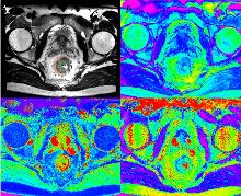
图1
基于MAGiC生成的合成图像以及肿瘤和瘤周脂肪的勾画A:合成T2WI图像,绿色区域为肿瘤ROI,红色区域为瘤周脂肪ROI;B:合成T1RT mapping 图像;C:合成T2RT mapping 图像;D:合成PD mapping图像。
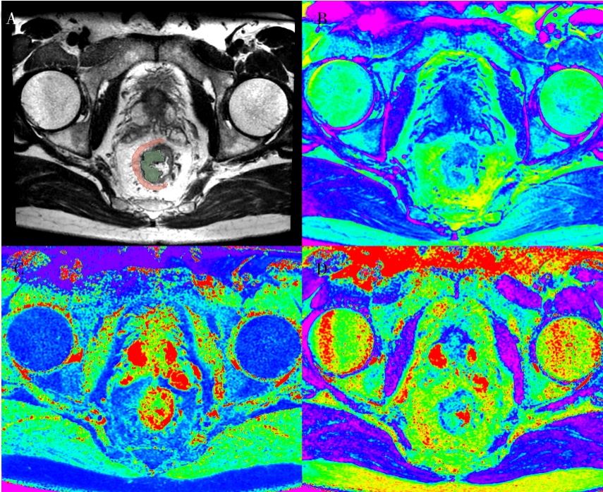
表2
各量化参数一致性检验
| ROI | ICC | P10 | P90 | Entropy | IQR | Mean | Median | Kurtosis | Maximum | MAD | Min | RMS | RMAD | Uniformity | |
|---|---|---|---|---|---|---|---|---|---|---|---|---|---|---|---|
| T1RT | Fat | intra | 0.990 | 0.788 | 0.950 | 0.953 | 0.949 | 0.984 | 0.763 | 0.801 | 0.831 | 0.901 | 0.897 | 0.937 | 0.958 |
| inter | 0.971 | 0.840 | 0.927 | 0.865 | 0.918 | 0.946 | 0.658 | 0.643 | 0.807 | 0.751 | 0.874 | 0.866 | 0.949 | ||
| Tumor | intra | 0.987 | 0.991 | 0.972 | 0.987 | 0.991 | 0.993 | 0.931 | 0.934 | 0.986 | 0.833 | 0.990 | 0.994 | 0.967 | |
| inter | 0.857 | 0.982 | 0.928 | 0.983 | 0.976 | 0.982 | 0.827 | 0.867 | 0.970 | 0.589 | 0.978 | 0.990 | 0.936 | ||
| T2RT | Fat | intra | 0.935 | 0.982 | 0.894 | 0.948 | 0.831 | 0.972 | 0.991 | 0.927 | 0.968 | 0.690 | 0.973 | 0.958 | 0.958 |
| inter | 0.901 | 0.904 | 0.894 | 0.905 | 0.863 | 0.958 | 0.977 | 0.905 | 0.930 | 0.618 | 0.913 | 0.920 | 0.939 | ||
| Tumor | intra | 0.987 | 0.990 | 0.973 | 0.965 | 0.993 | 0.995 | 0.844 | 0.839 | 0.971 | 0.946 | 0.993 | 0.974 | 0.977 | |
| inter | 0.954 | 0.948 | 0.833 | 0.903 | 0.969 | 0.977 | 0.524 | 0.721 | 0.872 | 0.793 | 0.966 | 0.906 | 0.902 | ||
| PD | Fat | intra | 0.951 | 0.984 | 0.894 | 0.927 | 0.984 | 0.984 | 0.851 | 0.956 | 0.911 | 0.797 | 0.985 | 0.920 | 0.900 |
| inter | 0.959 | 0.985 | 0.901 | 0.922 | 0.985 | 0.976 | 0.768 | 0.932 | 0.915 | 0.613 | 0.987 | 0.925 | 0.906 | ||
| Tumor | intra | 0.994 | 0.996 | 0.982 | 0.977 | 0.997 | 0.997 | 0.774 | 0.954 | 0.977 | 0.917 | 0.997 | 0.988 | 0.986 | |
| inter | 0.982 | 0.978 | 0.906 | 0.911 | 0.987 | 0.988 | 0.424 | 0.945 | 0.911 | 0.688 | 0.987 | 0.940 | 0.921 |
表3
ypN+与ypN-组间量化参数的差异性检验
| Quantitative parameter | Post-CRT pN stage | P | |
|---|---|---|---|
| 0 | 1-2 | ||
| T2RT_Fat_P90 | 129.60 (125.50, 135.00) | 139.53 (131.28, 144.48) | 0.025 |
| T2RT_Fat_RMS | 115.48 ± 7.16 | 120.09 ± 7.22 | 0.033 |
| T2RT_Fat_Maximum | 174.00 (150.70, 188.20) | 189.18 (165.71, 230.32) | 0.036 |
| T2RT_Fat_Uniformity | 0.61 ± 0.12 | 0.54 ± 0.10 | 0.035 |
表4
各量化参数及mrEMVI状态预测nCRT后ypN状态的ROC检验
| Quantitative parameter | AUC | Sensitivity | Specificity | PPV | NPV | Cutoff |
|---|---|---|---|---|---|---|
| T2RT_Fat_Maximum | 0.680 | 0.944 | 0.364 | 0.447 | 0.923 | 154.30 |
| T2RT_Fat_Uniformity | 0.673 | 0.889 | 0.121 | 0.356 | 0.667 | 0.437 |
| T2RT_Fat_P90 | 0.692 | 0.778 | 0.606 | 0.519 | 0.833 | 131.15 |
| T2RT_Fat_RMS | 0.678 | 0.611 | 0.788 | 0.611 | 0.788 | 119.95 |
| mrEMVI | 0.667 | 0.667 | 0.667 | 0.522 | 0.786 | / |
| Model | 0.747 | 0.778 | 0.667 | 0.560 | 0.846 | 0.32 |
表5
T2RT_Fat_P90,mrEMVI和逻辑回归模型预测效能的差异性检验
| Quantitative parameter | T2RT_Fat_P90 | mrEMVI | Model | |
|---|---|---|---|---|
| T2RT_Fat_P90 | \ | 0.808 | 0.260 | |
| mrEMVI | 0.808 | \ | 0.710 | |
| Model | 0.260 | 0.710 | \ | |
| [1] | SUNG H, FERLAY J, SIEGEL R L, et al. Global Cancer Statistics 2020: GLOBOCAN estimates of incidence and mortality worldwide for 36 cancers in 185 countries[J]. CA Cancer J Clin,2021,71(3): 209-249. |
| [2] | 范伯男, 李岩. 全球主要疾病负担及健康状况趋势分析——1990年至2021年全球疾病和伤害负担报告解读 [J]. 诊断学理论与实践,2024,23(5):474-483. |
| FAN B, LI Y. Trends in global major disease burden and health conditions—interpretation of the Global Burden of Disease Study 1990-2021[J]. J Diagn Concepts Pract, 2024,23(5):474-483. | |
| [3] | BENSON A B, VENOOK A P, ADAM M, et al. Colon Cancer, Version 3.2024, NCCN Clinical Practice Guidelines in Oncology [J]. J Natl Compr Canc Netw,2024,22(2 D). |
| [4] | HABR-GAMA A, PEREZ R O, NADALIN W, et al. Operative versus nonoperative treatment for stage 0 distal rectal cancer following chemoradiation therapy: long-term results[J]. Ann Surg,2004,240(4):711-717; discussion 7-8. |
| [5] |
MAAS M, NELEMANS P J, VALENTINI V, et al. Long-term outcome in patients with a pathological complete response after chemoradiation for rectal cancer: a pooled analysis of individual patient data[J]. Lancet Oncol,2010,11(9):835-44.
doi: 10.1016/S1470-2045(10)70172-8 pmid: 20692872 |
| [6] |
PAHLMAN L, BOHE M, CEDERMARK B, et al. The Swedish rectal cancer registry[J]. Br J Surg,2007,94(10):1285-1292.
pmid: 17661309 |
| [7] | TULCHINSKY H, SHMUELI E, FIGER A, et al. An interval >7 weeks between neoadjuvant therapy and surgery improves pathologic complete response and disease-free survival in patients with locally advanced rectal cancer [J]. Ann Surg Oncol,2008,15(10):2661-2667. |
| [8] |
VAN DER PAARDT M P, ZAGERS M B, BEETS-TAN R G, et al. Patients who undergo preoperative chemoradiotherapy for locally advanced rectal cancer restaged by using diagnostic MR imaging: a systematic review and meta-analysis [J]. Radiology,2013,269(1):101-112.
doi: 10.1148/radiol.13122833 pmid: 23801777 |
| [9] |
SEO N, KIM H, CHO M S, et al. Response assessment with MRI after chemoradiotherapy in rectal cancer: current evidences[J]. Korean J Radiol,2019,20(7):1003-1018.
doi: 10.3348/kjr.2018.0611 pmid: 31270972 |
| [10] | ZHANG S, YU M, CHEN D, et al. Role of MRI-based radiomics in locally advanced rectal cancer (Review)[J]. Oncol Rep,2022,47(2):34. |
| [11] |
RAFAELSEN S R, VAGN-HANSEN C, SORENSEN T, et al. Ultrasound elastography in patients with rectal cancer treated with chemoradiation[J]. Eur J Radiol,2013,82(6):913-917.
doi: 10.1016/j.ejrad.2012.12.030 pmid: 23410908 |
| [12] | HAGIWARA A, FUJIMOTO K, KAMAGATA K, et al. Age-related changes in relaxation times, proton density, myelin, and tissue volumes in adult brain analyzed by 2-dimensional quantitative synthetic magnetic resonance imaging[J]. Invest Radiol,2021,56(3):163-172. |
| [13] |
WARNTJES J B, LEINHARD O D, WEST J, et al. Rapid magnetic resonance quantification on the brain: optimization for clinical usage[J]. Magn Reson Med,2008,60(2): 320-329.
doi: 10.1002/mrm.21635 pmid: 18666127 |
| [14] |
KUMAR N M, FRITZ B, STERN S E, et al. Synthetic MRI of the knee: phantom validation and comparison with conventional MRI[J]. Radiology,2018,289(2):465-477.
doi: 10.1148/radiol.2018173007 pmid: 30152739 |
| [15] |
CUI Y, HAN S, LIU M, et al. Diagnosis and grading of prostate cancer by relaxation maps from synthetic MRI [J]. J Magn Reson Imaging,2020,52(2):552-564.
doi: 10.1002/jmri.27075 pmid: 32027071 |
| [16] | LIAN S, LIU H, MENG T, et al. Quantitative synthetic MRI for predicting locally advanced rectal cancer response to neoadjuvant chemoradiotherapy[J]. Eur Radiol,2023,33(3):1737-1745. |
| [17] |
SMITH N J, SHIHAB O, ARNAOUT A, et al. MRI for detection of extramural vascular invasion in rectal cancer[J]. Am J Roentgenol,2008,191(5):1517-1522.
doi: 10.2214/AJR.08.1298 pmid: 18941094 |
| [18] | ZHANG L N, XIAO W W, XI S Y, et al. Pathological Assessment of the AJCC tumor regression grading system after preoperative chemoradiotherapy for chinese locally advanced rectal cancer[J]. Medicine (Baltimore),2016,95(3):e2272. |
| [19] |
RINGEL A E, DRIJVERS J M, BAKER G J, et al. Obesity shapes metabolism in the tumor microenvironment to suppress anti-tumor immunity[J]. Cell,2020,183(7):1848-1866.
doi: 10.1016/j.cell.2020.11.009 pmid: 33301708 |
| [20] | JENSEN M D. Visceral fat: culprit or canary? [J]. Endocrinol Metab Clin North Am,2020,49(2):229-237. |
| [21] | LINO-SILVA L S, SALCEDO-HERNANDEZ R A, GAMBOA-DOMINGUEZ A. Tumour budding in rectal cancer. A comprehensive review[J]. Contemp Oncol (Pozn),2018,22(2):61-74. |
| [22] | 多中心直肠癌真实世界数据库建设与数据质量控制策略[J]. 中华消化外科杂志,2025,24(1):77-81. |
| Multicenter rectal cancer real-world database construction and data quality control strategies[J]. Chin Dig Surg, 2025,24(1):77-81. | |
| [23] | 李珂璇, 肖体先, 汪晓东, 等. 中低位直肠癌初诊及新辅助治疗后评估完成度分析:全国多中心真实世界研究[J]. 中华消化外科杂志,2025,24(1):113-119. |
| LI K X, XIAO T X, WANG X D, et al. Analysis of completion rate of tumor evaluation at initial assessment and after neoadjuvant therapy for mid and low rectal cancer : a national multicenter real-world study[J]. Chin J Dig Surg,2025,24(1):113-119. | |
| [24] | 毛翌皓, 冯青阳, 许剑民. 人工智能时代机器人结直肠癌手术的现状及进展[J]. 中华消化外科杂志, 2024, 23(4): 573-578. |
| MAO Y H, FENG Q Y, XU J M. Current status and progress of robotic colorectal cancer surgery in the era of artificial intelli-gence[J]. Chin J Dig Surg,2024,23(4):573-578. | |
| [25] |
SHIROUZU K, ISOMOTO H, KAKEGAWA T, et al. A prospective clinicopathologic study of venous invasion in colorectal cancer[J]. Am J Surg,1991,162(3):216-222.
pmid: 1928581 |
| [26] |
KRASNA M J, FLANCBAUM L, CODY R P, et al. Vascular and neural invasion in colorectal carcinoma. Incidence and prognostic significance[J]. Cancer,1988,61(5):1018-1023.
pmid: 3338045 |
| [1] | 张慧慧, 方姝, 吴梦雄, 刘方韬, 贺娜英, 董海鹏, 严福华. 深度学习重建技术在改善磁共振冠状位T1WI显示垂体神经内分泌肿瘤图像质量中的研究[J]. 诊断学理论与实践, 2024, 23(06): 594-601. |
| [2] | 查云飞, 武夏夏. MRI深度学习图像重建技术在肌骨系统疾病诊断的应用进展[J]. 诊断学理论与实践, 2024, 23(02): 114-118. |
| [3] | 高梦, 柴维敏, 严福华. 胰腺囊性肿瘤的CT/MRI诊断进展[J]. 诊断学理论与实践, 2024, 23(02): 184-191. |
| [4] | 李明, 陈克敏, 潘自来, 罗禹. CT及MRI预测急性缺血性脑梗死出血性转化的价值研究进展[J]. 诊断学理论与实践, 2024, 23(01): 83-89. |
| [5] | 丁景峰, 敖炜群, 朱珍, 孙静, 徐良根, 郑世保, 俞晶晶, 胡金文. 基于T2WI和DWI的磁共振影像组学在术前预测直肠癌壁外血管侵犯的价值研究[J]. 诊断学理论与实践, 2024, 23(01): 46-56. |
| [6] | 周熠磊, 张淼, 郭睿, 周金鑫, 李彪, 李翔. 18F-PSMA PET/MRI在早期诊断前列腺癌根治术后复发、转移中的价值[J]. 诊断学理论与实践, 2023, 22(06): 567-572. |
| [7] | 秦晓丹, 孙慧玲, 潘蓓, 潘玉琴, 王书奎. miR-1229-3p抑制结直肠癌疾病进展及作为潜在生物标志物的研究[J]. 诊断学理论与实践, 2023, 22(05): 429-440. |
| [8] | 冯丽, 任刚, 蔡嵘, 汪心韵, 王辉, 祝明洁. 泌尿生殖系统血管周上皮样细胞瘤(PEComa)的临床特征分析[J]. 诊断学理论与实践, 2023, 22(05): 460-465. |
| [9] | 李笑石, 秦越. 影像学技术在痛风诊断及疾病监测中的应用研究进展[J]. 诊断学理论与实践, 2023, 22(03): 311-318. |
| [10] | 李卫侠, 徐学勤, 朱晓雷, 陈克敏. 39例肾上腺皮质癌患者的CT、MRI影像特点及其诊断价值[J]. 诊断学理论与实践, 2023, 22(02): 134-140. |
| [11] | 陈乾, 林慧敏, 严福华. 磁共振成像评估肝功能储备的研究进展[J]. 诊断学理论与实践, 2023, 22(02): 190-196. |
| [12] | 李佳曦, 汪锦江, 俞立萍, 袁英, 乔光磊, 马俐君. RAB25沉默抑制结直肠癌细胞铁死亡的作用研究[J]. 诊断学理论与实践, 2022, 21(06): 710-718. |
| [13] | 杨蕊馨, 杜宇童, 燕然林, 朱正纲, 李琛, 于颖彦. 消化道肿瘤单细胞转录组测序研究中生物样本前处理改良的探索[J]. 诊断学理论与实践, 2022, 21(05): 567-574. |
| [14] | 王文涵, 夏蜀珺, 詹维伟. 长链非编码RNA ENST00000489676在超声评估甲状腺乳头状癌颈部淋巴结转移中的应用[J]. 诊断学理论与实践, 2022, 21(04): 514-519. |
| [15] | 徐琛莹, 李嫣然, 倪晓枫, 徐上妍, 林青. 超声预测老年甲状腺乳头状癌患者颈部淋巴结转移的效能及相关超声征象分析[J]. 诊断学理论与实践, 2022, 21(03): 343-348. |
| 阅读次数 | ||||||
|
全文 |
|
|||||
|
摘要 |
|
|||||