Journal of Diagnostics Concepts & Practice ›› 2024, Vol. 23 ›› Issue (05): 494-499.doi: 10.16150/j.1671-2870.2024.05.005
• Original articles • Previous Articles Next Articles
LI Ying1,2( ), JIANG Han1,2, WANG Xiaoxue1,2, WEI Haonan1,2
Received:2024-03-20
Accepted:2024-08-08
Online:2024-10-25
Published:2025-02-25
Contact:
LI Ying
E-mail:liying@ihcams.ac.cn
CLC Number:
LI Ying, JIANG Han, WANG Xiaoxue, WEI Haonan. Analysis of chest CT findings, diagnosis, and treatment of mucormycosis infection in 65 hematologic disease patients[J]. Journal of Diagnostics Concepts & Practice, 2024, 23(05): 494-499.
Table 1
Types of chest CT findings
| Thoracic CT manifestations | Cases | Reversed halo syndrome | Halo syndrome | Pleural effusion |
|---|---|---|---|---|
| Single large patchy consolidation | 23(35%) | 18(78%) | 0 | 10(43%) |
| Multiple nodules | 28(43%) | 0 | 25(89%) | 20(71%) |
| Mixed | 11(17%) | 8(73%) | 0 | 2(18%) |
| Diffuse exudative type | 3(5%) | 3(100%) | 0 | 2(67%) |

Figure 1
Mucormycotic foci in lung A: Female, 40 years old, with acute myeloid leukemia, undergoing chemotherapy, presented with cough and reduced sputum, without fever; chest CT revealed consolidation in the right middle lobe with reverse halo sign, presenting as a single large patchy consolidation; B and C: Male, 48 years old, asymptomatic after acute myeloid leukemia transplantation. B showed thickening of the vessels in the posterior basal segment of the left lower lobe, while C revealed the lesion progressed to mass-like consolidation after 6 days,and enhanced chest CT revealed local vessel cut-off sign, presenting as a single large patchy consolidation; D: Female, 22 years old, with chest pain after chemotherapy for acute myeloid leukemia; chest CT scan revealed multiple nodules with halo sign in the left upper lobe, presenting as a multiple nodule pattern; E and F: Female, 53 years old, post-transplantation for acute lymphoblastic leukemia, with low-grade fever, cough, and breathlessness;E represented multiple scattered patchy and nodular shadows in both lungs; After 10 days, the lesion area increased and there was a large consolidation in the upper lobe of the left lung with a halo sign (F).
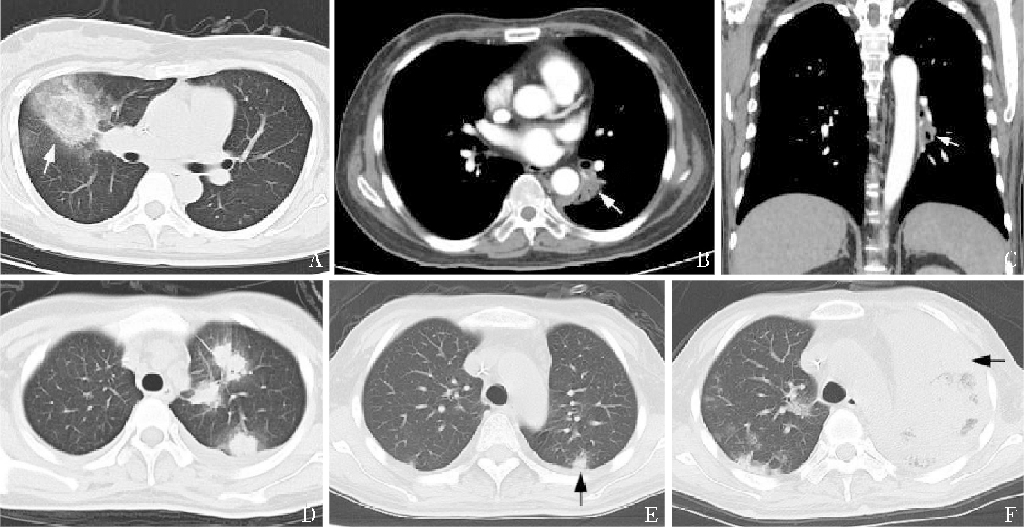

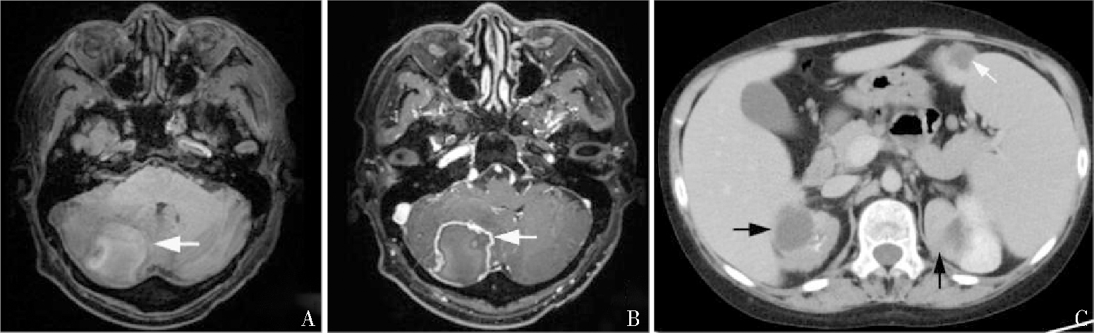
Figure 2
Mucormycotic foci in brain, kidney and spleen A, B, C showed mucormycotic lesions in the brain, kidney and spleen, respectively.
| [1] | VALENTINE J C, MORRISSEY C O, TACEY M A, et al. A population-based analysis of attributable hospitalisation costs of invasive fungal diseases in haematological malignancy patients using data linkage of state-wide regi-stry and costing databases: 2009-2015[J]. Mycoses, 2020, 63(2):162-171. |
| [2] | PETRIKKOS G, SKIADA A, LORTHOLARY O, et al. Epidemiology and clinical manifestations of mucormycosis[J]. Clin Infect Dis, 2012,54 Suppl 1:S23-34. |
| [3] | YANG N, ZHANG L, FENG S. Clinical features and treatment progress of invasive mucormycosis in patients with hematological malignancies[J]. J Fungi (Basel), 2023, 9(5):592. |
| [4] |
DANION F, AGUILAR C, CATHERINOT E, et al. Mucormycosis: new developments into a persistently devastating infection. Semin Respir Crit Care Med, 2015, 36(5):692-705.
doi: 10.1055/s-0035-1562896 pmid: 26398536 |
| [5] | 齐瑶, 赵明峰, 邓琦, 等. 血液病合并毛霉菌病七例报道并文献复习[J]. 中华血液学杂志, 2019, 40(11):943-947. |
| QI Y, ZHAO M F, DENG Q, et al. Report of seven cases of hematological diseases combined with trichoderma and literature review[J]. Chin J Hematol, 2019, 40(11):943-947. | |
| [6] | 中国医药教育协会真菌病专业委员会, 中国毛霉病专家共识工作组. 中国毛霉病临床诊疗专家共识(2022)[J]. 中华内科杂志, 2023, 62(6):597-605. |
| Fungal Disease Professional Committee of China Medical Education Association, Expert Consensus Working Group on Trichoderma in China. Consensus of clinical diagnosis and treatment experts on trichoderma in China (2022)[J]. Chin J Intern Med, 2023, 62(6):597-605. | |
| [7] | 吴挺, 周华, 顾海艇, 等. 肺毛霉菌病25例临床高危因素特征及预后分析[J]. 中华医学杂志, 2018, 98(32):2579-2582. |
| WU T, ZHOU H, GU H T, et al. Clinical high-risk factors and prognosis analysis of 25 cases of pulmonary mucormycosis[J]. Natl Med J China, 2018, 98(32):2579-2582. | |
| [8] |
AGRAWAL R, YELDANDI A, SAVAS H, et al. Pulmonary mucormycosis: risk factors, radiologic findings, and pathologic correlation[J]. Radiographics, 2020, 40(3):656-666.
doi: 10.1148/rg.2020190156 pmid: 32196429 |
| [9] | 许尚文, 陈自谦, 钟群, 等. 肺毛霉菌病的CT及正电子发射计算机体层成像-CT表现[J]. 中华放射学杂志, 2014, 48(7):551-554. |
| XU S W, CHEN Z Q, ZHONG Q, et al. CT and positron emission computed tomography CT manifestations of pulmonary mucormycosis[J]. Chin J Radiol, 2014, 48(7):551-554. | |
| [10] | HAMMER M M, MADAN R, HATABU H. Pulmonary mucormycosis: radiologic features at presentation and over time[J]. AJR Am J Roentgenol., 2018, 210(4):742-747. |
| [11] | JUNG J, KIM M Y, LEE H J, et al. Comparison of computed tomographic findings in pulmonary mucormycosis and invasive pulmonary aspergillosis[J]. Clin Microbiol Infect, 2015, 21(7):684.e11-684.e18. |
| [12] |
BOURCIER J, HEUDES P M, MORIO F, et al. Prevalence of the reversed halo sign in neutropenic patients compared with non-neutropenic patients: Data from a single-centre study involving 27 patients with pulmonary mucormycosis (2003-2016)[J]. Mycoses, 2017, 60(8):526-533.
doi: 10.1111/myc.12624 pmid: 28429890 |
| [13] | 石玉铸, 王鹏飞, 宋杰, 等. 白血病造血干细胞移植后肺部毛霉菌病CT特征表现[J]. 实用放射学杂志, 2017, 33(6):851-853. |
| SHI Y Z, WANG P F, SONG J, et al. CT features of pulmonary mucormycosis after leukemia hematopoietic stem cell transplantation[J]. J Pract Radiol, 2017, 33(6):851-853. | |
| [14] |
CHAMILOS G, MAROM E M, LEWIS R E, et al. Predictors of pulmonary zygomycosis versus invasive pulmonary aspergillosis in patients with cancer[J]. Clin Infect Dis, 2005, 41(1):60-66.
doi: 10.1086/430710 pmid: 15937764 |
| [15] |
NAM B D, KIM T J, LEE K S, et al. Pulmonary mucormycosis: serial morphologic changes on computed tomography correlate with clinical and pathologic findings[J]. Eur Radiol, 2018, 28(2):788-795.
doi: 10.1007/s00330-017-5007-5 pmid: 28812135 |
| [16] | 周红俐, 刘范林, 宋兰. 肺毛霉菌病的CT表现及动态随诊[J]. 实用放射学杂志, 2020, 36(8): 1216-1219. |
| ZHOU H L, LIU F L, SONG L. CT manifestations and dynamic follow-up of pulmonary mucormycosis[J]. J Pract Radiol, 2020, 36(8): 1216-1219. | |
| [17] | LERSY F, ROYER-LEBLOND J, LHERMITTE B, et al. Cerebral mucormycosis: neuroimaging findings and histopathological correlation[J]. J Neurol, 2022, 269(3):1386-1395. |
| [18] |
KHABA M C, NEVONDO L M, MOROATSHEHLA S M, et al. Disseminated mucormycosis presenting as a renal mass in an human immunodeficiency virus-infected patient: a case report[J]. S Afr J Infect Dis, 2021, 36(1):202.
doi: 10.4102/sajid.v36i1.202 pmid: 34485490 |
| [1] | HUANG Ruikun, YANG Yanzhao, CHAI Weimin. Advances in application of photon-counting CT for pancreatic imaging [J]. Journal of Diagnostics Concepts & Practice, 2025, 24(02): 111-117. |
| [2] | LI Weixia, YAN Fuhua. Photon-counting CT in liver disease: applications and advances [J]. Journal of Diagnostics Concepts & Practice, 2025, 24(02): 118-124. |
| [3] | WANG Mengzhen, BAO Shouyu, LIU Peng, YAN Fuhua, YANG Wenjie. Application of photon-counting CT in cardiovascular diseases [J]. Journal of Diagnostics Concepts & Practice, 2025, 24(02): 125-134. |
| [4] | CHANG Rui, LI Jiqiang, YANG Yanzhao, CHAI Weimin, YAN Fuhua, DONG Haipeng.. Evaluation value of single-phase images from photon-counting CT-based low-dose pancreatic dynamic volume perfusion scanning for pancreatic cancer imaging [J]. Journal of Diagnostics Concepts & Practice, 2025, 24(02): 155-162. |
| [5] | LI Zhuohan, HUANG Xinyun, GUO Rui, LI Biao. 18F-FDG PET/CT in the diagnosis and prognosis evaluation of follicular lymphoma [J]. Journal of Diagnostics Concepts & Practice, 2024, 23(04): 439-444. |
| [6] | GAO Meng, CHAI Weimin, YAN Fuhua. Advance in study on diagnosis of pancreatic cystic tumors on CT/MRI imaging [J]. Journal of Diagnostics Concepts & Practice, 2024, 23(02): 184-191. |
| [7] | LI Ming, CHEN Kemin, PAN Zilai, LUO Yu. Research progress on the value of CT and MRI in predicting hemorrhagic transformation after acute ischemic stroke [J]. Journal of Diagnostics Concepts & Practice, 2024, 23(01): 83-89. |
| [8] | ZHOU Yilei, ZHANG Miao, GUO Rui, ZHOU Jinxin, LI Biao, LI Xiang. Value of 18F-PSMA PET/MRI for early diagnosis of recurrence and metastasis in prostate cancer patients after radical prostatectomy [J]. Journal of Diagnostics Concepts & Practice, 2023, 22(06): 567-572. |
| [9] | FENG Li, REN Gang, CAI Rong, WANG Xinyun, WANG Hui, ZHU Mingjie. Clinical features study of perivascular epithelioid cell tumor (PEComa) in genitourinary system [J]. Journal of Diagnostics Concepts & Practice, 2023, 22(05): 460-465. |
| [10] | DONG Lai, WANG Wei, WU Jialiang, LIU Yanpu, GUAN Xin, CHEN Kemin. Pulmonary imaging manifestations and related research progress of lymphangioleiomyomatosis [J]. Journal of Diagnostics Concepts & Practice, 2023, 22(05): 501-506. |
| [11] | YANG Wenjie, YAN Fuhua. Interpretation of the Clinical Practice Guidelines for Lung Cancer Screening (version 2) of 2022 National Comprehensive Cancer Network(NCCN) [J]. Journal of Diagnostics Concepts & Practice, 2023, 22(01): 14-20. |
| [12] | XU Hao, ZHANG Zhi, XIE Xueqian, YANG Wenyi, LIU Shaowen. Comparative study on software DEEPVESSEL FFR and invasive FFR in assessing coronary ischemia [J]. Journal of Diagnostics Concepts & Practice, 2021, 20(04): 384-390. |
| [13] | CAO Qiqi, QIN Le, ZHOU Huijuan, YANG Zhitao, SU Wenting, YANG Wenjie, CHENG Zenghui, LU Yong, YAN Fuhua, PAN Zilai. CT features of 2019 novel coronavirus pneumonia [J]. Journal of Diagnostics Concepts & Practice, 2020, 19(1): 16-19. |
| [14] | WANG Lan, ZHANG Huan, GE Yingqian, LU Jing, CUI Zheng, YAN Ling, PAN Zilai. Clinical application and evaluation of artificial intelligence-assisted semi-automatic segmentation software for detection of liver metastases from gastric cancer: intra-observer and inter-observer differences [J]. Journal of Diagnostics Concepts & Practice, 2019, 18(05): 515-520. |
| [15] | HU Meng, ZHU Huaishi, SONG Qi. Low dose muti-slice computed tomography in preoperative evaluation of location for dental implantation procedure [J]. Journal of Diagnostics Concepts & Practice, 2018, 17(01): 66-69. |
| Viewed | ||||||
|
Full text |
|
|||||
|
Abstract |
|
|||||