Journal of Diagnostics Concepts & Practice ›› 2023, Vol. 22 ›› Issue (05): 460-465.doi: 10.16150/j.1671-2870.2023.05.007
• Original articles • Previous Articles Next Articles
FENG Li1a,2, REN Gang1a( ), CAI Rong3(), WANG Xinyun1a, WANG Hui2, ZHU Mingjie1b
Received:2022-05-08
Online:2023-10-25
Published:2024-03-15
CLC Number:
FENG Li, REN Gang, CAI Rong, WANG Xinyun, WANG Hui, ZHU Mingjie. Clinical features study of perivascular epithelioid cell tumor (PEComa) in genitourinary system[J]. Journal of Diagnostics Concepts & Practice, 2023, 22(05): 460-465.
Table 1
Clinical and imaging findings of 5 cases of perivascular epithelioid cell tumor of genitourinary system
| Serial numb-er | Gen-der | Age (years old) | Symptom | NSE | Location | Shape | Volume(mm) | Density | Necrosis | Enhancement type | Enhanced degree | Small blood vessels travel after enhanc-ement | Renal venous cancer thromb-us | Distant metastasis of tumor |
|---|---|---|---|---|---|---|---|---|---|---|---|---|---|---|
| No.1 | male | sixty | No symptoms | high | Right kidney | Round | 9×7×8 | Homog-eneous | - | Fast in and out | Obvious | - | - | - |
| No.2 | male | nine | Abdominal mass | high | Left kidney | Irregular | 150×160×190 | Heterog-eneous | + | Fast in and slow out | Medium | + | - | - |
| No.3 | fema-le | sixty-one | No symptom | Norm-al | Right kidney | Oval | 100×161×190 | Heterog-eneous | - | Fast in and slow out | Medium | - | - | - |
| No.4 | male | sixty-three | Ventosity | Norm-al | Left kidney | Irregular | 50×60×25 | Heterog-eneous | + | Fast in and slow out | Medium | + | - | - |
| No.5 | fema-le | nine | Vaginal fluid | high | Vagina | Round | 21×17×25 | Homog-eneous | - | Enhancement appearances | Obvious | - | - | - |
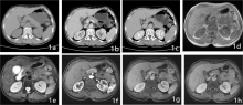
Figure 1
PEComa in the right kidney (Case 1) 1a-1c: axial, a small nodular lesions in the right kidney grows outward with clear boundaries. CT value of plain scan is 46 HU, and CT values of the arterial and venous phases after enhancement are 101 HU and 76 HU respectively, showing fast in and out enhancement. 1a shows slightly high density on plain scan, 1b shows obvious enhancement in the arterial phase. 1c shows obvious decrease in venous phase and low density relatively. 1d-1h: a small nodular lesion of the right kidney growing outward, equal signal on T1WI and slightly low signal on T2WI, obviously enhancement in arterial phase, decreased enhancement in venous phase and delayed phase, showing relatively low signal.

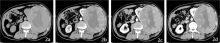
Figure 2
PEComa in the left kidney (Case 2) 2a: axial, a large mixed density lesion of the left kidney is found in plain scan, with a low density area in the center and surrounding solid density. 2b: obvious enhancement of the surrounding solid part of the arterial phase, with vascular shadow visible inside, but no enhancement in the central low density area, and the lesion invading the psoas major and retroperitoneal space. 2b: the solid part of the focus in the venous phase continued to enhance significantly, but no enhancement was observed in the central low-density area. 2c-2d: the solid part of the focus in the delayed phase decreased in enhancement, but no enhancement was observed in the central low-density area.
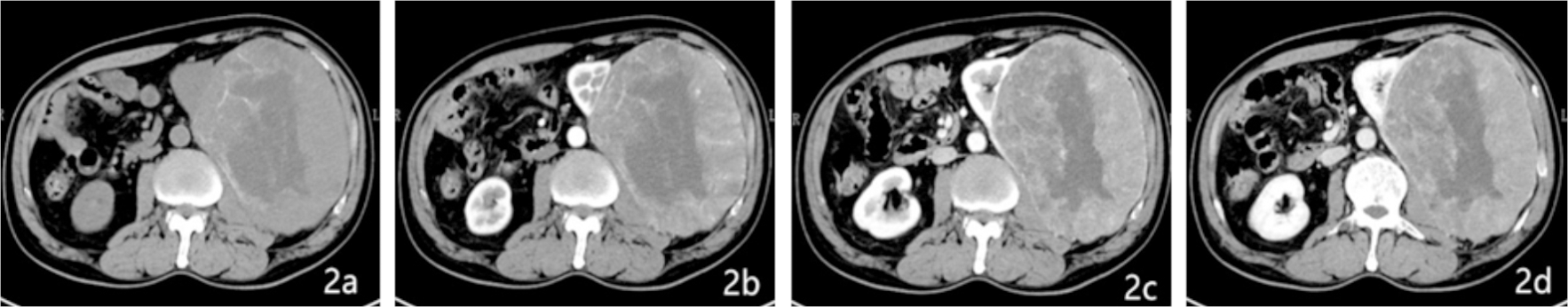
Figure 3
PEComa in the vagina (Case 3) 3a: the right lateral wall of the lower vaginal segment was local thickening and distention in CT plain scan, and a small isodensity nodule was observed, with homogeneous density. 3b: obvious enhancement at arterial phase, 3c: decreased enhancement at venous phase.

Table 2
Pathologic results of 5cases of perivascular epithelioid cell tumor of genitourinary system
| Serial number | HMB45 | MelanA | SMA | CD31 | CD34 | S-100 |
|---|---|---|---|---|---|---|
| NO.1 | + | - | - | - | + | - |
| NO.2 | + | + | - | - | - | + |
| NO.3 | + | - | + | - | - | - |
| NO.4 | + | - | - | - | + | + |
| NO.5 | + | - | - | - | - | - |
| [1] |
PEA M, BONETTI F, ZAMBONI G, et al. Clear cell tumor and angiomyolipoma[J]. Am J Surg Pathol, 1991, 15(2):199-202.
pmid: 2025321 |
| [2] |
BONETTI F, PEA M, MARTIGNONI G, et al. Clear cell("sugar") tumor of the lung is a lesion strictly related to angiomyolipoma--the concept of a family of lesions characterized by the presence of the perivascular epithelioid cells (PEC)[J]. Pathology, 1994, 26(3):230-236.
doi: 10.1080/00313029400169561 URL |
| [3] |
MARTIGNONI G, PEA M, ZAMPINI C, et al. PEComas of the kidney and of the genitourinary tract[J]. Semin Diagn Pathol, 2015, 32(2):140-159.
doi: 10.1053/j.semdp.2015.02.006 pmid: 25804448 |
| [4] |
ZAMBONI G, PEA M, MARTIGNONI G, et al. Clear cell "sugar" tumor of the pancreas. A novel member of the family of lesions characterized by the presence of perivascular epithelioid cells[J]. Am J Surg Pathol, 1996, 20(6):722-730.
doi: 10.1097/00000478-199606000-00010 pmid: 8651352 |
| [5] |
FOLPE A L, GOODMAN Z D, ISHAK K G, et al. Clear cell myomelanocytic tumor of the falciform ligament/ligamentum teres: a novel member of the perivascular epithelioid clear cell family of tumors with a predilection for children and young adults[J]. Am J Surg Pathol, 2000, 24(9):1239-1246.
doi: 10.1097/00000478-200009000-00007 pmid: 10976698 |
| [6] |
THWAY K, FISHER C. PEComa: morphology and gene-tics of a complex tumor family[J]. Ann Diagn Pathol, 2015, 19(5)359-368.
doi: 10.1016/j.anndiagpath.2015.06.003 URL |
| [7] |
MARTIGNONI G, PEA M, REGHELLIN D, et al. PEComas: the past, the present and the future[J]. Virchows Arch, 2008, 452(2):119-132.
doi: 10.1007/s00428-007-0509-1 pmid: 18080139 |
| [8] | 吴小鹏, 陈静, 张力峰, 等. 肾血管周上皮样细胞肿瘤一例报告[J]. 中华泌尿外科杂志, 2013, 34(8):607. |
| WU X P, CHEN J, ZHANG L F, et al. Renal perivascular epithelioid cell tumor a case report[J]. Chin J Urol, 2013, 34(8)607. |
| [1] | DONG Lai, WANG Wei, WU Jialiang, LIU Yanpu, GUAN Xin, CHEN Kemin. Pulmonary imaging manifestations and related research progress of lymphangioleiomyomatosis [J]. Journal of Diagnostics Concepts & Practice, 2023, 22(05): 501-506. |
| [2] | LI Xiaoshi, QIN Yue. Multiple radiology imaging techniques in the diagnosis of gout [J]. Journal of Diagnostics Concepts & Practice, 2023, 22(03): 311-318. |
| [3] | CHEN Qian, LIN Huimin, YAN Fuhua. Advances in the evaluation of hepatic function by magnetic resonance imaging [J]. Journal of Diagnostics Concepts & Practice, 2023, 22(02): 190-196. |
| [4] | YANG Wenjie, YAN Fuhua. Interpretation of the Clinical Practice Guidelines for Lung Cancer Screening (version 2) of 2022 National Comprehensive Cancer Network(NCCN) [J]. Journal of Diagnostics Concepts & Practice, 2023, 22(01): 14-20. |
| [5] | HUANG Juan, ZHU Xiaolei, LI Xiao, CHEN Kemin, YAN Fuhua, XU Xueqin. Study on blood oxygen level-dependent magnetic resonance imaging for the assessment of early renal hypoxia in chronic kidney disease [J]. Journal of Diagnostics Concepts & Practice, 2022, 21(03): 385-389. |
| [6] | ZHU Naiyi, JIANG Yixin, CHAI Li, CHAI Weimin. Diagnostic values of magnetic resonance imaging in mammography detected BI-RADS≥4 category calcifications with negative ultrasound results [J]. Journal of Diagnostics Concepts & Practice, 2021, 20(05): 439-444. |
| [7] | XU Hao, ZHANG Zhi, XIE Xueqian, YANG Wenyi, LIU Shaowen. Comparative study on software DEEPVESSEL FFR and invasive FFR in assessing coronary ischemia [J]. Journal of Diagnostics Concepts & Practice, 2021, 20(04): 384-390. |
| [8] | ZHANG Xuekun, LI Yan, YAN Fuhua, ZHAO Hongfei, SONG Qi. Application value of new accelerating technology based on constellation shuttling imaging in brain MRI [J]. Journal of Diagnostics Concepts & Practice, 2021, 20(04): 378-383. |
| [9] | SUN Tiantian, YE Baoying, YANG Yu, NIU Jianmei. Color Doppler ultrasound and magnetic resonance imaging in prenatal diagnosis of pernicious placenta previa and pernicious placenta previa with placenta accreta: clinic value and analysis of missed diagnosis [J]. Journal of Diagnostics Concepts & Practice, 2021, 20(02): 173-177. |
| [10] | CAO Juntao, HU Ming, QIAN Pingkang, TU Jianchun, ZHANG Huan, SHEN Junkang. Application value of 3.0T MRI 3D-MERGE sequence in evaluating the degree of supraspinatus tendon injury [J]. Journal of Diagnostics Concepts & Practice, 2021, 20(01): 77-81. |
| [11] | CAO Qiqi, QIN Le, ZHOU Huijuan, YANG Zhitao, SU Wenting, YANG Wenjie, CHENG Zenghui, LU Yong, YAN Fuhua, PAN Zilai. CT features of 2019 novel coronavirus pneumonia [J]. Journal of Diagnostics Concepts & Practice, 2020, 19(1): 16-19. |
| [12] | GU Xiaohong, SUN Aimin, WANG Qian, ZHU Ming, ZHONG Yumin. The three-dimensional balanced steady state free precession magnetic resonance imaging sequence in diagnosis of anomalous origin of the coronary artery from the pulmonary artery in children [J]. Journal of Diagnostics Concepts & Practice, 2020, 19(02): 145-150. |
| [13] | WU Shuang, XIE Qian, GUAN Xueni, ZHANG Sufang, GAO Xinfang, LIANG Zonghui. Perfomence of MRI intravoxel incoherent motion diffusion weighted imaging parameters in diagnosing active Crohn's disease [J]. Journal of Diagnostics Concepts & Practice, 2020, 19(02): 157-161. |
| [14] | CHEN Jie, HU Jin, YANG Kang, FU Yi. Analysis of risk factors and prognosis of cerebral hemorrhage patients accompanied by cortical superficial siderosis [J]. Journal of Diagnostics Concepts & Practice, 2019, 18(2): 133-138. |
| [15] | WANG Lan, ZHANG Huan, GE Yingqian, LU Jing, CUI Zheng, YAN Ling, PAN Zilai. Clinical application and evaluation of artificial intelligence-assisted semi-automatic segmentation software for detection of liver metastases from gastric cancer: intra-observer and inter-observer differences [J]. Journal of Diagnostics Concepts & Practice, 2019, 18(05): 515-520. |
| Viewed | ||||||
|
Full text |
|
|||||
|
Abstract |
|
|||||