Journal of Surgery Concepts & Practice ›› 2024, Vol. 29 ›› Issue (01): 67-73.doi: 10.16139/j.1007-9610.2024.01.11
• Review • Previous Articles Next Articles
WANG Ting, WANG Chaofu, YUAN Fei( )
)
Received:2024-01-10
Online:2024-01-25
Published:2024-05-14
Contact:
YUAN Fei
E-mail:daphny2014@163.com
CLC Number:
WANG Ting, WANG Chaofu, YUAN Fei. Progress in pathological diagnosis of intraductal papillary mucinous neoplasm of the pancreas[J]. Journal of Surgery Concepts & Practice, 2024, 29(01): 67-73.

Fig 1
Pathology gross photography of IPMN with invasive carcinoma
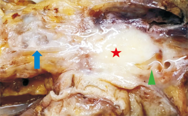

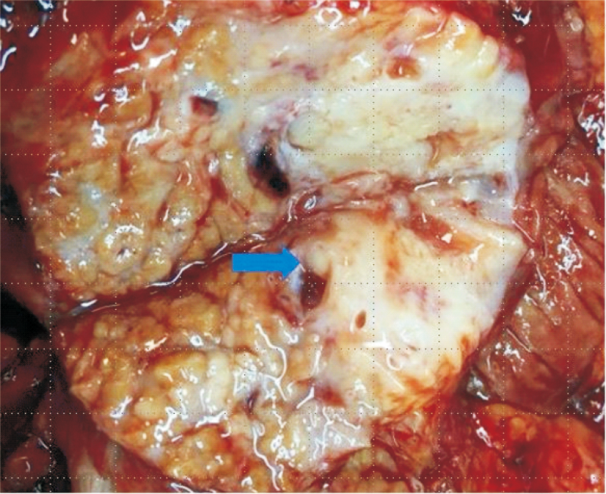
Fig 2
Pathology gross photography of IPMN with a mural nodule
Tab 1
Morphological characteristics of IPMN dysplassia
| 异型增生 级别 | 组织形态 | 细胞形态 | 核分裂象 |
|---|---|---|---|
| 低 | 肿瘤细胞排列成单层或具有轴心的乳头状结构 | 细胞轻-中度异型,细胞核位于基底部或呈假复层排列 | 罕见或偶见 |
| 高 | 肿瘤细胞排列成复杂分支乳头、筛孔状、出芽 | 细胞高度异型,细胞核多形性,缺乏极性 | 多见或靠近管腔表面 |
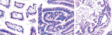
Fig 3
Pathological morphology of three differentiations of IPMN (HE staining, ×200)

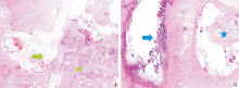
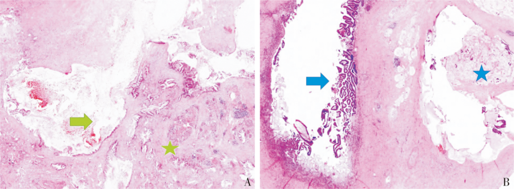
Fig 4
Pathological morphology of IPMN with invasive carcinoma (HE staining, ×100)
Tab 2
Histological and immunohistochemical characteristics of 5 types of pancreatic tumors
| IPMN | PanIN | MCN | ITPN | IOPN | |
|---|---|---|---|---|---|
| 组织学特征 | |||||
| 与胰管关系 | 相通 | 相通 | 不相通 | 相通 | 相通 |
| 附壁结节 | 有或无 | 无 | 无 | 有 | 有 |
| 上皮异型性 | 低级别、高级别 | 低级别、高级别 | 低级别、高级别 | 高级别 | 高级别 |
| 卵巢样间质 | 无 | 无 | 有 | 无 | 无 |
| 免疫组织化学特征 | |||||
| CK7/CK8/CK18/CK19 | + | + | + | + | + |
| CK20/MUC2/CDX2 | 肠型+ | - | - | - | 杯状细胞+ |
| MUC1 | 胰胆管型+ | + | -/+a) | + | + |
| MUC5AC | + | + | + | - | + |
| MUC6 | 可+,可- | 低级别+ | - | + | + |
| 其他特殊标记 | 无 | 无 | 有b) | 无 | 有c) |

Fig 5
Pathological morphology of PanIN (HE staining, ×200)
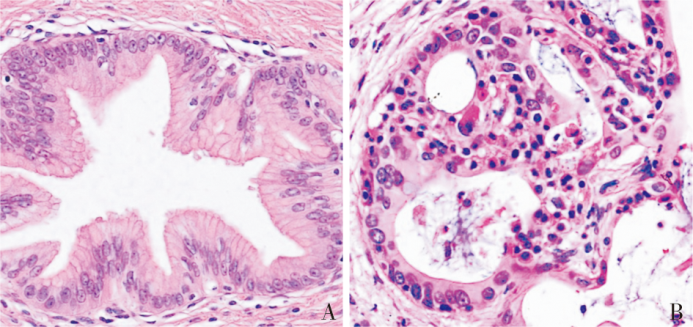
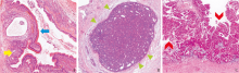
Fig 6
Pathological morphology of three pancreatic tumors that need to be distinguished from IPMN (HE staining, ×100)

| [1] |
NAGTEGAAL I D, ODZE R D, KLIMSTRA D, et al. The 2019 WHO classification of tumours of the digestive system[J]. Histopathology, 2020, 76(2):182-188.
doi: 10.1111/his.13975 pmid: 31433515 |
| [2] | WOOD L D, ADSAY N V, BASTURK O, et al. Syste-matic review of challenging issues in pathology of intraductal papillary mucinous neoplasms[J]. Pancreatology, 2023, 23(7):878-891. |
| [3] | HRUBAN R H, KLIMSTRA D S, ZAMBONI G, et al. A semicentennial of pancreatic pathology: the genetic revolution is here, but don't throw the baby out with the bath water![J]. Hum Pathol, 2020,95:99-112. |
| [4] |
COMPAGNO J, OERTEL J E. Microcystic adenomas of the pancreas (glycogen-rich cystadenomas): a clinicopathologic study of 34 cases[J]. Am J Clin Pathol, 1978, 69(3):289-298.
doi: 10.1093/ajcp/69.1.289 pmid: 637043 |
| [5] |
PARAMYTHIOTIS D, KARLAFTI E, FOTIADOU G, et al. Pancreatic intraductal papillary mucinous neoplasms: a narrative review[J]. Acta Med Litu, 2023, 30(1):53-65.
doi: 10.15388/Amed.2023.30.1.6 pmid: 37575378 |
| [6] |
ZAMBONI G, SCARPA A, BOGINA G, et al. Mucinous cystic tumors of the pancreas: clinicopathological features, prognosis, and relationship to other mucinous cystic tumors[J]. Am J Surg Pathol, 1999, 23(4):410-422.
pmid: 10199470 |
| [7] | ASSARZADEGAN N, BABANIAMANSOUR S, SHI J. Updates in the diagnosis of intraductal neoplasms of the pancreas[J]. Front Physiol, 2022,13:856803. |
| [8] |
TANAKA M, FERNÁNDEZ-DEL CASTILLO C, KAMISAWA T, et al. Revisions of international consensus Fukuoka guidelines for the management of IPMN of the pancreas[J]. Pancreatology, 2017, 17(5):738-753.
doi: S1424-3903(17)30516-1 pmid: 28735806 |
| [9] |
ATTIYEH M A, FERNÁNDEZ-DEL CASTILLO C, AL EFISHAT M, et al. Development and validation of a multi-institutional preoperative nomogram for predicting grade of dysplasia in intraductal papillary mucinous neoplasms (ipmns) of the pancreas: a report from the pancreatic surgery consortium[J]. Ann Surg, 2018, 267(1):157-163.
doi: 10.1097/SLA.0000000000002015 pmid: 28079542 |
| [10] |
VEGE S S, ZIRING B, JAIN R, et al. American gastroenterological association institute guideline on the diagnosis and management of asymptomatic neoplastic pancreatic cysts[J]. Gastroenterology, 2015, 148(4):819-822.
doi: 10.1053/j.gastro.2015.01.015 pmid: 25805375 |
| [11] |
ELTA G H, ENESTVEDT B K, SAUER B G, et al. ACG clinical guideline: diagnosis and management of pancreatic cysts[J]. Am J Gastroenterol, 2018, 113(4):464-479.
doi: 10.1038/ajg.2018.14 pmid: 29485131 |
| [12] |
European Study Group on Cystic Tumours of the Pancreas. European evidence-based guidelines on pancreatic cystic neoplasms[J]. Gut, 2018, 67(5):789-804.
doi: 10.1136/gutjnl-2018-316027 pmid: 29574408 |
| [13] | LEE J E, CHOI S Y, MIN J H, et al. Determining malignant potential of intraductal papillary mucinous neoplasm of the pancreas: CT versus MRI by using revised 2017 international consensus guidelines[J]. Radiology, 2019, 293(1):134-143. |
| [14] |
D'ONOFRIO M, TEDESCO G, CARDOBI N, et al. Magnetic resonance (MR) for mural nodule detection studying Intraductal papillary mucinous neoplasms (IPMN) of pancreas: imaging-pathologic correlation[J]. Pancreatology, 2021, 21(1):180-187.
doi: 10.1016/j.pan.2020.11.024 pmid: 33376061 |
| [15] |
CHON H K, SONG T J, YOO K H, et al. Enhancing mural nodules in the main pancreatic duct of main and mixed types of intraductal papillary mucinous neoplasms: does size matter in malignancy risk?[J]. Gut Liver, 2023, 17(6):942-948.
doi: 10.5009/gnl220378 pmid: 37317514 |
| [16] | ASSARZADEGAN N, THOMPSON E, SALIMIAN K, et al. Pathology of intraductal papillary mucinous neoplasms[J]. Langenbecks Arch Surg, 2021, 406(8):2643-2655. |
| [17] | TRIANTOPOULOU C, GOURTSOYIANNI S, KARAKAXAS D, et al. Intraductal papillary mucinous neoplasm of the pancreas: a challenging diagnosis[J]. Diagnostics (Basel), 2023, 13(12):2015. |
| [18] | KAWAKAMI Y, KOSHITA S, KANNO Y, et al. Pancreatic ductal adenocarcinomas concomitant with intraductal papillary mucinous neoplasms of the pancreas: a investigation of those clinicopathological features by analyzing 48 patients who underwent surgery for those lesions[J]. Pancreatology, 2023, 23(6):674-681. |
| [19] | HASHIMOTO D, SATOI S, YAMAMOTO T, et al. Long-term outcomes of patients with multifocal intraductal papillary mucinous neoplasm following pancreatectomy[J]. Pancreatology, 2022, 22(7):1046-1053. |
| [20] |
KOBAYASHI T, OMORI Y, ONO Y, et al. Pathways for the development of multiple epithelial types of intraductal papillary mucinous neoplasm of the pancreas[J]. J Gastroenterol, 2021, 56(6):581-592.
doi: 10.1007/s00535-021-01783-2 pmid: 33796937 |
| [21] | NOË M, BROSENS L A A. Gastric- and intestinal-type IPMN: two of a kind?[J]. Virchows Arch, 2020, 477(1):17-19. |
| [22] | AMINI N, HABIB J R, BLAIR A, et al. Invasive and noninvasive progression after resection of noninvasive intraductal papillary mucinous neoplasms[J]. Ann Surg, 2022, 276(2):370-377. |
| [23] | REZAEE N, BARBON C, ZAKI A, et al. Intraductal pa-pillary mucinous neoplasm (IPMN) with high-grade dysplasia is a risk factor for the subsequent development of pancreatic ductal adenocarcinoma[J]. HPB (Oxford), 2016, 18(3):236-246. |
| [24] |
MAJUMDER S, PHILIP N A, SINGH NAGPAL S J, et al. High-Grade dysplasia in resected Main-Duct Intraductal Papillary Mucinous Neoplasm (MD-IPMN) is associated with an increased risk of subsequent pancreatic cancer[J]. Am J Gastroenterol, 2019, 114(3):524-529.
doi: 10.1038/s41395-018-0403-2 pmid: 30413822 |
| [25] |
BASTURK O, HONG S M, WOOD L D, et al. A revised classification system and recommendations from the baltimore consensus meeting for neoplastic precursor lesions in the pancreas[J]. Am J Surg Pathol, 2015, 39(12):1730-1741.
doi: 10.1097/PAS.0000000000000533 pmid: 26559377 |
| [26] | OHTSUKA T, FERNANDEZ-DEL CASTILLO C, FURUKAWA T, et al. International evidence-based Kyoto guidelines for the management of intraductal papillary mucinous neoplasm of the pancreas[J]. Pancreatology, 2024, 24(2):255-270. |
| [27] | PFLÜGER M J, GRIFFIN J F, HACKENG W M, et al. The impact of clinical and pathological features on intraductal papillary mucinous neoplasm recurrence after surgical resection: long-term follow-up analysis[J]. Ann Surg, 2022, 275(6):1165-1174. |
| [28] | LEONHARDT C S, HINZ U, KAISER J, et al. Presence of low-grade IPMN at the pancreatic transection margin does not have prognostic significance after resection of IPMN-associated pancreatic adenocarcinoma[J]. Eur J Surg Oncol, 2023, 49(1):113-121. |
| [29] |
DHAR V K, MERCHANT N B, PATEL S H, et al. Does surgical margin impact recurrence in noninvasive intraductal papillary mucinous neoplasms?: a multi-institutional study[J]. Ann Surg, 2018, 268(3):469-478.
doi: 10.1097/SLA.0000000000002923 pmid: 30063495 |
| [30] | OMORI Y, FURUKAWA T, SCARPA A, et al. Co-occurring IPMN and pancreatic cancer: the same or different? An overview from histology to molecular pathology[J]. J Clin Pathol, 2023, 76(11):734-739. |
| [31] |
SOTOZONO H, KANKI A, YASOKAWA K, et al. Value of 3-T MR imaging in intraductal papillary mucinous neoplasm with a concomitant invasive carcinoma[J]. Eur Radiol, 2022, 32(12):8276-8284.
doi: 10.1007/s00330-022-08881-6 pmid: 35665843 |
| [32] | YAMAGUCHI K, KANEMITSU S, HATORI T, et al. Pancreatic ductal adenocarcinoma derived from IPMN and pancreatic ductal adenocarcinoma concomitant with IPMN[J]. Pancreas, 2011, 40(4):571-580. |
| [33] | IYER M K, SHI C, ECKHOFF A M, et al. Digital spatial profiling of intraductal papillary mucinous neoplasms: toward a molecular framework for risk stratification[J]. Sci Adv, 2023, 9(11):eade4582. |
| [34] |
OMORI Y, ONO Y, TANINO M, et al. Pathways of progression from intraductal papillary mucinous neoplasm to pancreatic ductal adenocarcinoma based on molecular features[J]. Gastroenterology, 2019, 156(3):647-661.
doi: S0016-5085(18)35160-6 pmid: 30342036 |
| [35] |
FELSENSTEIN M, NOË M, MASICA D L, et al. IPMNs with co-occurring invasive cancers: neighbours but not always relatives[J]. Gut, 2018, 67(9):1652-1662.
doi: 10.1136/gutjnl-2017-315062 pmid: 29500184 |
| [36] |
FISCHER C G, BELEVA GUTHRIE V, BRAXTON A M, et al. Intraductal papillary mucinous neoplasms arise from multiple independent clones, each with distinct mutations[J]. Gastroenterology, 2019, 157(4):1123-1137.
doi: S0016-5085(19)40987-6 pmid: 31175866 |
| [37] | PEA A, YU J, REZAEE N, et al. Targeted DNA sequen-cing reveals patterns of local progression in the pancreatic remnant following resection of Intraductal Papillary Mucinous Neoplasm (IPMN) of the pancreas[J]. Ann Surg, 2017, 266(1):133-141. |
| [38] |
KIM H, RO J Y. Intraductal tubulopapillary neoplasm of the pancreas: an overview[J]. Arch Pathol Lab Med, 2018, 142(3):420-423.
doi: 10.5858/arpa.2016-0405-RSR2 pmid: 29494224 |
| [39] | ITOH T, OMORI Y, SEINO M, et al. Gene Rearrangement and expression of prkaca and prkacb govern morphobiology of pancreatobiliary oncocytic neoplasms[J]. Mod Pathol, 2024, 37(1):100358. |
| [40] |
INNOCENTI L, ROTONDO M I, DONATI F, et al. Intraductal oncocytic papillary neoplasm (IOPN): two case reports and review of the literature[J]. Transl Cancer Res, 2023, 12(3):663-672.
doi: 10.21037/tcr-22-2029 pmid: 37033351 |
| [1] | FENG Meijing, REN Xinping, ZHAN Weiwei, ZHENG Lili, LI Junjian. Value of contrast-enhanced ultrasound in differentiating benign and malignant gallbladder lesions which diameter more than 1 cm [J]. Journal of Surgery Concepts & Practice, 2023, 28(06): 556-562. |
| [2] | LI Xiaoshi, QIN Yue. Multiple radiology imaging techniques in the diagnosis of gout [J]. Journal of Diagnostics Concepts & Practice, 2023, 22(03): 311-318. |
| [3] | YANG Qiao, FU Xin, WANG Zhe, LIU Tantan. Cytopathologic analysis of thyroid secondary tumors [J]. Journal of Diagnostics Concepts & Practice, 2023, 22(03): 270-276. |
| [4] | WU Nanming, LI Jun, TAO Juan. Hot spots in diagnosis of malignant melanoma [J]. Journal of Diagnostics Concepts & Practice, 2023, 22(03): 215-220. |
| [5] | XIE Yaqiong, LIN Xiaoyi. Value of serum-free light chain assay in differential diagnosis and staging of nephropathy of various etiologies [J]. Journal of Diagnostics Concepts & Practice, 2023, 22(02): 166-171. |
| [6] | HAO Jiaqi, WANG Xinlu, HU Xiaofan, PAN Xiaoxia, XU Jing, MA Jun. Clinical differential diagnosis of acute tubulointerstitial nephritis and acute tubular necrosis [J]. Journal of Diagnostics Concepts & Practice, 2023, 22(02): 127-133. |
| [7] | LUO Fangxiu, MA Qianchen, YUAN Fei. The fifth edition of WHO classification of digestive system tumors: update and progress on biliary system tumors [J]. Journal of Surgery Concepts & Practice, 2023, 28(02): 124-131. |
| [8] | WANG Zhaohui, WU Haibo. Clinicopathological analysis of 31 cases of gastric schwannoma [J]. Journal of Diagnostics Concepts & Practice, 2021, 20(06): 552-556. |
| [9] | WANG Jianjun, CHEN Ya, FAN Xiangshan, NIU Fengnan. Sclerosing angiomatoid nodular transformation of spleen: clinicopathological analysis and literature review [J]. Journal of Diagnostics Concepts & Practice, 2019, 18(05): 560-564. |
| [10] | CHANG Rui, XU Jiaxu, DONG Haipeng, WU Mengxiong, ZHAO Xuesong, MIAO Fei, YAN Fuhua. Value of CT spectral imaging in the evaluation of Crohn's disease activity [J]. Journal of Diagnostics Concepts & Practice, 2019, 18(04): 432-435. |
| [11] | YANG Ruxue, LI Nan, ZHOU Ting, ZHAO Yan, CHEN Shaohua, ZHU Qing, FENG Zhenzhong. Clinicopathologic analysis of skin melanocyte lesions [J]. Journal of Diagnostics Concepts & Practice, 2018, 17(05): 566-571. |
| [12] | WU Xinyang, ZHANG Huan, PAN Zilai, TAN Jingwen, GAO Xiaoyuan. The diagnostic value of dual-source CT in differentiating primary gastric lymphoma from advanced gastric cancer [J]. Journal of Diagnostics Concepts & Practice, 2018, 17(01): 60-65. |
| [13] | XU Haimin, ZHANG Peipei. Comparison of performance between three types of automatic immunohistochemical stainer in pathological diagnosis of breast cancer [J]. Journal of Diagnostics Concepts & Practice, 2017, 16(06): 645-649. |
| [14] | ZHU Peipei, ZOU Jue, CHEN Jun, XU Rongrong, YAN Hongzhu. Intracranial solitary fibrous tumor/hemangiopericytoma: a clinicopathologic study of 20 cases with review of literature [J]. Journal of Diagnostics Concepts & Practice, 2017, 16(06): 622-626. |
| [15] | YI Lin, XIAO Li, CHEN Yan, YIN Yulei. Anaplastic large cell lymphoma: a clinicopathological study and review of literature [J]. Journal of Diagnostics Concepts & Practice, 2017, 16(03): 313-319. |
| Viewed | ||||||
|
Full text |
|
|||||
|
Abstract |
|
|||||